Silent heart attack: What are the risks?

A silent heart attack, medically termed a “silent myocardial infarction,” is a potentially dangerous event that often goes unnoticed due to its lack of typical symptoms. Unlike a traditional heart attack, which is accompanied by chest pain, shortness of breath, and discomfort in the arms, neck, jaw, or back, a silent heart attack occurs without these apparent signs. This makes it a significant concern, as individuals are less likely to seek immediate medical attention, leading to delayed diagnosis and treatment. Understanding the risks associated with silent heart attacks is crucial for prevention and proactive healthcare management.

Causes and Risks

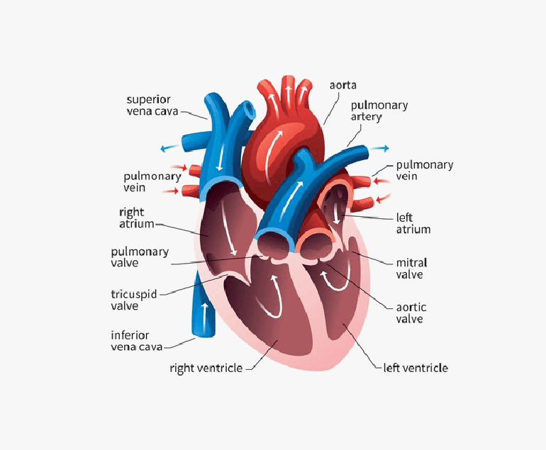
Silent heart attacks share the same underlying cause as symptomatic heart attacks: a disruption of blood flow to a portion of the heart muscle due to a blocked or narrowed coronary artery. This interruption leads to the death of heart muscle cells, which can impair cardiac function and increase the risk of future heart-related complications. The factors that contribute to silent heart attacks are often the same as those for traditional heart attacks, including high blood pressure, high cholesterol, diabetes, obesity, smoking, and a sedentary lifestyle. However, certain demographic groups, such as older adults and individuals with diabetes, are more prone to experiencing silent heart attacks.

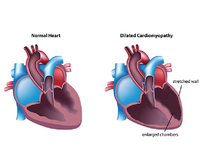
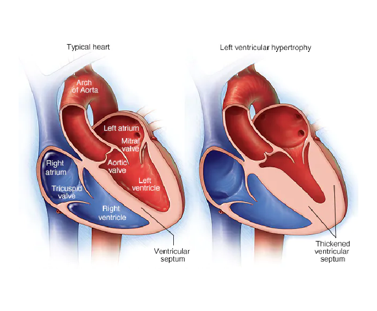
One of the major dangers of silent heart attacks is their gradual progression. Without the characteristic chest pain that typically prompts individuals to seek medical help, silent heart attacks can go undetected for weeks, months, or even years. During this time, untreated heart damage may accumulate, putting individuals at greater risk of heart failure, abnormal heart rhythms (arrhythmias), and subsequent heart attacks. Additionally, silent heart attacks are often identified incidentally when a person undergoes medical tests for unrelated issues, further highlighting the hidden nature of these events.

Preventive Measures

The lack of awareness surrounding silent heart attacks highlights the importance of preventive healthcare measures. Regular check-ups and screenings are essential, especially for individuals with risk factors. Blood pressure, cholesterol, and blood sugar levels should be closely monitored, and lifestyle modifications should be adopted to mitigate these risk factors. Adopting a heart-healthy diet rich in fruits, vegetables, whole grains, lean proteins, and healthy fats, along with engaging in regular physical activity, can significantly reduce the likelihood of both silent and symptomatic heart attacks.

Moreover, recognizing potential warning signs that might not be as dramatic as traditional symptoms is vital. Unexplained fatigue, mild discomfort in the chest, nausea, or discomfort in the upper abdomen, back, or jaw could all be indicative of a silent heart attack. Hence, they should not be ignored, especially if they persist or worsen over time.

Conclusion

Silent heart attacks pose a grave risk due to their inconspicuous nature and delayed diagnosis. The absence of classic symptoms can lead to untreated heart damage and increase the likelihood of complications. It is essential for individuals, particularly those with risk factors, to prioritize regular health check-ups, maintain a heart-healthy lifestyle, and be vigilant about any unusual or persistent symptoms. By taking proactive measures, individuals can mitigate the risks associated with silent heart attacks and promote overall heart health.

Cardiac Amyloidosis

Cardiac amyloidosis is a rare and potentially fatal disorder that arises from the abnormal accumulation of amyloid protein in the heart tissues, leading to compromised cardiac function.

Nature of Cardiac Amyloidosis

Cardiac amyloidosis stems from the buildup of amyloid fibrils, which are insoluble protein aggregates that accumulate in various organs, including the heart. These amyloid deposits infiltrate the heart muscles, causing them to become stiff, thickened, and less compliant. Over time, this impairs the heart’s ability to pump blood effectively, leading to heart failure and other cardiovascular complications.

Subtypes of Cardiac Amyloidosis

There are different subtypes of cardiac amyloidosis, each originating from distinct precursor proteins. The two most prevalent subtypes are:

  • AL Amyloidosis (Immunoglobulin Light Chain Amyloidosis): This form arises when abnormal plasma cells in the bone marrow produce excessive amounts of light chains, a component of antibodies. These light chains then accumulate as amyloid fibrils in various organs, including the heart.
  • ATTR Amyloidosis (Transthyretin Amyloidosis): In ATTR amyloidosis, the precursor protein is transthyretin, a protein responsible for transporting thyroid hormone and retinol. Mutations or aging-related changes in transthyretin can lead to the formation of amyloid fibrils that deposit in the heart.

Challenges in Diagnosis

Cardiac amyloidosis remains a diagnostic challenge due to its overlapping symptoms with other heart conditions and its rarity. Common symptoms include fatigue, shortness of breath, swelling in the legs, and irregular heartbeats. These nonspecific signs often delay accurate diagnosis, which is further complicated by the fact that cardiac amyloidosis can coexist with other heart diseases.

Advancements in Detection

Recent advancements in imaging techniques have greatly improved the detection of cardiac amyloidosis. Cardiac MRI, nuclear imaging with bone tracers, and echocardiography can provide insights into the presence and extent of amyloid deposits in the heart. Additionally, advances in genetic testing help in identifying ATTR amyloidosis, enabling early intervention for individuals with genetic predisposition.

Management and Treatment

Managing cardiac amyloidosis involves addressing both the underlying cause and the associated symptoms. In AL amyloidosis, treatments focus on suppressing the abnormal plasma cells responsible for producing the amyloidogenic light chains. Chemotherapy and stem cell transplantation are commonly used in the treatment. For ATTR amyloidosis, treatment options include stabilizing the transthyretin protein or, in cases of hereditary ATTR, gene-silencing therapies.

Heart Disease and Pericardiocentesis

Heart disease is one of the leading causes of mortality and requires advanced medical interventions. Pericardiocentesis is a critical procedure in addressing complications related to pericardial effusion – a condition characterized by the accumulation of fluid in the pericardium, the protective sac around the heart. This procedure plays a vital role in both diagnosing and treating underlying heart conditions, alleviating symptoms, and preventing potentially life-threatening complications.

Causes

Pericardial effusion can result from various factors such as infections, inflammation, malignancies, trauma, or as a consequence of cardiac surgery. The accumulation of excess fluid within the pericardial space can exert pressure on the heart, impairing its ability to pump blood effectively. This can lead to symptoms like chest pain, shortness of breath, and even cardiac tamponade – a condition where the pressure from the fluid buildup compresses the heart chambers, compromising its function and circulation.

Procedure

Pericardiocentesis involves the insertion of a needle or catheter into the pericardial space to drain the accumulated fluid. This procedure serves a dual purpose: providing symptomatic relief by relieving the pressure on the heart and facilitating diagnostic evaluation by analyzing the removed fluid for potential underlying causes.

The pericardiocentesis procedure is performed under imaging guidance, such as echocardiography or fluoroscopy, to ensure precision and minimize the risk of complications. The needle is carefully guided into the pericardial sac, and the fluid is aspirated. In some cases, a catheter may be left in place for continuous drainage if there is a significant fluid buildup or if the effusion is recurrent.

Benefits

Pericardiocentesis can provide rapid relief from the debilitating symptoms caused by pericardial effusion. By promptly reducing the pressure on the heart, patients often experience a marked improvement in their ability to breathe and reduced discomfort. Additionally, the removed fluid can be sent for analysis, which helps in diagnosing the underlying cause of the effusion. This diagnostic insight is crucial in guiding subsequent treatment decisions.

In some cases, pericardiocentesis may serve as a temporary measure while other interventions, such as medication, are initiated to address the underlying condition causing the effusion. However, in more severe cases where pericardial effusion poses an immediate threat to cardiac function, pericardiocentesis can be a life-saving intervention.

Conclusion

Pericardiocentesis plays a vital role in the management of heart disease, specifically in addressing the complications arising from pericardial effusion. This procedure offers both diagnostic and therapeutic benefits, helping in the swift mitigation of symptoms and potentially preventing life-threatening cardiac complications.

Tricuspid Valve Disease

The tricuspid valve is an essential component of the heart and regulates blood flow between the right atrium and right ventricle. Tricuspid valve disease is a medical condition that affects the functionality of this valve, potentially leading to significant cardiovascular complications.

Categories

Tricuspid valve disease can be categorized into two main types: tricuspid regurgitation and tricuspid stenosis. Tricuspid regurgitation occurs when the valve doesn’t close properly, allowing blood to flow back into the right atrium during the ventricular contraction. This puts an extra strain on the heart and can lead to symptoms like fatigue, swelling in the legs and abdomen, and an enlarged liver.

On the other hand, tricuspid stenosis involves a narrowing of the valve, which restricts blood flow from the right atrium to the right ventricle. This condition can result in symptoms such as fatigue, palpitations, and fluid retention.

Causes

The causes of tricuspid valve disease can vary. It may be a result of congenital defects, where the valve doesn’t form properly during foetal development. Acquired tricuspid valve disease is often associated with other heart conditions, such as rheumatic fever, infective endocarditis, or pulmonary hypertension. These underlying conditions can damage the valve’s structure and function over time.

Diagnosis

Diagnosing tricuspid valve disease typically involves a combination of medical history assessment, physical examination, and various imaging techniques. Echocardiography is commonly used to visualize the valve’s structure and assess blood flow. Electrocardiograms (ECGs) and chest X-rays can help evaluate the heart’s electrical activity and overall condition.

Treatment

Treatment options for tricuspid valve disease depend on the severity of the condition and the associated symptoms. In mild cases, close monitoring and lifestyle modifications, such as reducing sodium intake and managing fluid retention, may be sufficient. For more severe cases, medical interventions or surgical procedures might be necessary.

Medical management often includes medications to alleviate symptoms and manage underlying conditions. Surgical interventions can range from minimally invasive techniques to open-heart surgery, depending on the complexity of the disease and the patient’s overall health.

Conclusion

Tricuspid valve disease is a complex cardiovascular condition that can have significant implications for a person’s health. Early detection and appropriate management are crucial to prevent the progression of the disease and the development of potentially life-threatening complications.

67-Year-Old Woman Gets Life as Doctors Create History with Dual Surgeries

Medical science has made significant strides in the field of surgery, shattering age barriers for medical interventions. In a significant achievement, a team of skilled doctors at BLK-Max Super Specialty Hospital successfully performed two surgeries on a 67-year-old woman, granting her a new lease of life. Dr. Ramji Mehrotra, the Chief of Cardiology & Vascular Surgery at BLK-Max Super Specialty Hospital, led the remarkable medical feat.

According to Dr. Mehrotra, the surgical team utilized bilateral mammary artery grafts for Coronary Artery Bypass surgery, demonstrating exceptional coordination. Given the complexity of the case, they opted for a safe myocardial revascularization with total arterial bypass, which yielded outstanding results. Arterial grafts are more likely to remain patent even without blood thinners.

Due to the dedicated efforts of Dr. Mehrotra and the medical team at BLK-Max Super Specialty Hospital, the patient made a swift recovery and was discharged in just six days after the surgery.

Ref: thehealthsite.com

Hypertrophic Cardiomyopathy

Hypertrophic cardiomyopathy (HOCM) is a complex and potentially life-threatening heart condition characterized by abnormal thickening (hypertrophy) of the heart muscle. It is one of the most common genetic heart disorders, affecting people of all ages and backgrounds.

Causes

Hypertrophic cardiomyopathy is primarily caused by mutations in genes responsible for the structure and function of the heart muscle. These genetic mutations lead to abnormal growth and arrangement of the cardiac muscle cells, resulting in the characteristic thickening of the heart muscle.

HOCM is usually inherited in an autosomal dominant pattern, meaning that a child has a 50% chance of inheriting the condition if one of the parents is affected. However, spontaneous gene mutations can also occur, leading to the development of HOCM in individuals without a family history of the condition.

Symptoms

The symptoms of hypertrophic cardiomyopathy can vary widely among individuals. Some may remain asymptomatic throughout their lives, while others may experience severe symptoms. Common symptoms include shortness of breath, chest pain, heart palpitations, fatigue, dizziness, fainting, and lightheadedness. These symptoms can worsen during physical exertion or strenuous activities and may be indicative of an increased risk of sudden cardiac arrest, especially in young athletes.

Diagnosis

The diagnosis of hypertrophic cardiomyopathy involves a comprehensive evaluation of the patient’s medical history, a physical examination, and a variety of diagnostic tests. These tests may include electrocardiography (ECG), echocardiography, cardiac MRI, exercise stress tests, and genetic testing. ECG can help identify abnormal electrical patterns in the heart, while echocardiography and cardiac MRI provide detailed images of the heart’s structure and function. Genetic testing is performed to identify specific gene mutations associated with HOCM.

Treatment

The management of hypertrophic cardiomyopathy focuses on relieving symptoms, preventing complications, and reducing the risk of sudden cardiac arrest. Treatment plans are tailored to individual patients based on their symptoms, risk factors, and disease progression. Medications such as beta-blockers, calcium channel blockers, and anti-arrhythmic drugs are commonly prescribed to control heart rate, rhythm, and blood pressure.

In some cases, surgical procedures like septal myectomy or alcohol septal ablation may be recommended to relieve obstruction of blood flow from the heart. Implantable cardioverter-defibrillators (ICDs) may also be used in high-risk patients to prevent sudden cardiac arrest.

Prognosis

The prognosis for individuals with hypertrophic cardiomyopathy varies depending on the severity of the disease, the presence of complications, and the effectiveness of treatment. Many people with HOCM lead normal lives with minimal symptoms and a low risk of complications. Regular monitoring, adherence to treatment plans, and lifestyle modifications, such as avoiding strenuous activities, can help improve outcomes and reduce the risk of complications.

Conclusion

Hypertrophic cardiomyopathy is a complex heart condition characterized by abnormal thickening of the heart muscle. It is primarily caused by genetic mutations and can present with a range of symptoms and complications. Early diagnosis, thorough evaluation, and personalized treatment plans are essential for managing HOCM effectively. With appropriate medical care and lifestyle modifications, individuals with hypertrophic cardiomyopathy can lead fulfilling lives while minimizing the risks associated with the condition.

Dilated Cardiomyopathy

Dilated cardiomyopathy (DCM) is a condition characterized by the dilation and weakening of the heart’s main pumping chamber, the left ventricle. This chronic and progressive disorder affects the heart muscle, leading to impaired cardiac function and potentially causing heart failure. DCM can also affect the right ventricle and both ventricles in some cases.

Causes

The exact causes of DCM are often unknown, but various factors can contribute to its development. These include genetic mutations, viral infections (such as viral myocarditis), exposure to toxins (for example alcohol), autoimmune diseases, and metabolic disorders. In many cases, DCM is considered idiopathic, meaning the cause is uncertain.

Symptoms

DCM can present with a range of symptoms, which may include fatigue, shortness of breath, swelling of the legs and ankles (edema), rapid or irregular heartbeat (arrhythmias), chest pain, dizziness, and fainting. These symptoms are often related to the heart’s inability to effectively pump blood and supply oxygen to the body’s tissues.

Diagnosis

To diagnose DCM, a comprehensive evaluation is required, which typically involves a thorough medical history review, physical examination, and various diagnostic tests. These tests may include electrocardiogram (ECG), echocardiogram (to assess heart structure and function), cardiac MRI or CT scan (for detailed imaging), blood tests (to rule out other conditions), and occasionally a heart biopsy.

Treatment

The management of DCM aims to improve symptoms, slow disease progression, and prevent complications. Treatment approaches may include:

  1. Medications: Several medications can be prescribed to manage symptoms, reduce the workload on the heart, control blood pressure, prevent blood clots, and treat arrhythmias. These may include beta-blockers, ACE inhibitors, diuretics, anticoagulants, and anti-arrhythmic drugs.
  • Lifestyle modifications: Patients need to make lifestyle changes like reducing salt intake, avoiding alcohol, stopping smoking, maintaining proper weight, and exercising as required.
  • Device therapy: In advanced cases or those with severe heart failure, implantable devices like pacemakers or defibrillators may be recommended to regulate the heart’s rhythm or deliver electrical shocks if life-threatening arrhythmias occur.
  • Cardiac rehabilitation: This supervised program combines exercise, education, and counseling to help patients recover and improve their overall cardiovascular health.
  • Surgical interventions: In some cases, surgical interventions may be necessary, such as coronary artery bypass grafting (CABG) to improve blood flow to the heart, or heart transplantation for end-stage heart failure.

Prognosis

The prognosis for individuals with DCM can vary widely depending on the underlying cause, severity of symptoms, and response to treatment. With appropriate management, lifestyle modifications, and adherence to medication, many patients can experience an improved quality of life and a slower disease progression. However, some individuals may develop more severe heart failure and require advanced therapies or heart transplantation. Regular follow-up with a cardiologist is essential to monitor the condition and adjust treatment as needed.

Conclusion

Dilated cardiomyopathy is a complex heart condition characterized by the enlargement and weakening of the heart muscle. While the exact causes are unclear in many cases, a combination of genetic, viral, autoimmune, and environmental factors can contribute to its development. Early diagnosis, appropriate medical management, lifestyle modifications, and sometimes surgical interventions are essential for managing the symptoms, slowing disease progression, and improving the prognosis for individuals with DCM.

Abdominal Aortic Aneurysm

Abdominal aortic aneurysm (AAA) is a potentially life-threatening condition characterized by the enlargement and weakening of the aorta—the main blood vessel that supplies oxygenated blood to the abdomen, pelvis, and lower extremities. AAA often develops slowly over time, and if left untreated, it can lead to a rupture, resulting in severe internal bleeding and potentially fatal consequences.

Causes

According to Dr. Ramji Mehrotra, India’s best heart surgeon, the exact cause of AAA is still unknown, but several risk factors have been identified. The primary risk factor is age, with individuals over 65 years being at higher risk. Other factors include smoking, high blood pressure, atherosclerosis (buildup of fatty deposits in the arteries), male gender, family history of AAA, and certain genetic disorders, such as Marfan syndrome and Ehlers-Danlos syndrome.

Symptoms

AAA often develops asymptomatically and is frequently detected incidentally during medical imaging tests conducted for unrelated conditions. However, as the aneurysm grows larger, it may cause symptoms such as:

  • Abdominal pain or discomfort: Dull, persistent pain or a pulsating sensation in the abdomen or back.
  • Swelling or a visible mass: A throbbing lump may be felt in the abdomen.
  • Rupture symptoms: Sudden and severe pain, dizziness, rapid heartbeat, low blood pressure, and loss of consciousness may occur if the aneurysm ruptures.

Diagnosis

AAA can be diagnosed through various imaging techniques, including:

  • Ultrasound: This non-invasive test uses sound waves to create images of the abdominal aorta, providing information about its size and structure.
  • Computed Tomography (CT) Scan: This imaging technique produces detailed cross-sectional images of the aorta, allowing for precise measurement and evaluation of the aneurysm.
  • Magnetic Resonance Imaging (MRI): MRI can provide additional information about the aorta and surrounding structures, especially in cases where CT scanning is contraindicated.

Treatment

The management of AAA depends on the size, growth rate, and overall health of the patient. Treatment options include:

  1. Watchful Waiting: Small aneurysms (less than 5.5 centimeters) that are not rapidly growing may be monitored regularly through imaging tests. Lifestyle modifications, such as quitting smoking and controlling blood pressure, are advised to reduce the risk of rupture.
  2. Surgery: For larger aneurysms or those growing rapidly, surgical intervention is often recommended. The two primary surgical procedures are open repair and endovascular aneurysm repair (EVAR).
  3. Open Repair: Involves surgically replacing the weakened section of the aorta with a synthetic graft.
  4. EVAR: A minimally invasive procedure where a stent graft is placed inside the aneurysm, providing support to the weakened vessel walls.

Conclusion

Dr. Ramji Mehrotra says that abdominal aortic aneurysm is a potentially life-threatening condition that requires careful monitoring and prompt management. Early diagnosis, regular screenings for at-risk individuals, and lifestyle modifications play a crucial role in preventing complications. It is important to consult with a doctor for accurate diagnosis, and appropriate treatment options.

Left Ventricle of the Heart

The left ventricle is a crucial component of the human heart, playing a vital role in pumping oxygenated blood to the rest of the body. It is one of the four chambers of the heart and is responsible for the majority of the heart’s pumping action.

Anatomy

The left ventricle is located at the bottom left side of the heart and is the largest and thickest chamber. It is responsible for receiving oxygen-rich blood from the left atrium and pumping it out to the body through the aorta, the largest artery in the body.

The walls of the left ventricle are highly muscular and contract forcefully during each heartbeat to propel blood into the circulation.

Function

The primary function of the left ventricle is to generate enough force to pump blood to the entire body. It achieves this by contracting and relaxing in a coordinated manner with the other chambers of the heart. During systole, the contraction phase, the left ventricle contracts forcefully, pushing blood into the aorta.

This blood is then distributed to various organs and tissues, delivering oxygen and nutrients essential for their functioning. In diastole, the relaxation phase, the left ventricle refills with blood from the left atrium, preparing for the next contraction.

Significance of the Left Ventricle

The left ventricle is often referred to as the “workhorse” of the heart due to its critical role in maintaining the circulation of oxygenated blood throughout the body. Its muscular walls are much thicker than those of the other chambers, enabling it to generate the necessary force for pumping blood against the high resistance of the systemic circulation. The left ventricle’s ability to contract forcefully is crucial for maintaining adequate blood flow and oxygen supply to the body’s organs and tissues.

In addition, the left ventricle is intricately connected to various aspects of cardiovascular health. It is commonly assessed through medical imaging techniques such as echocardiography and cardiac MRI to evaluate its size, shape, and function. Changes in the structure or function of the left ventricle can indicate underlying heart conditions, such as left ventricular hypertrophy, heart failure, or coronary artery disease. By monitoring the left ventricle’s health, medical professionals can diagnose and manage these conditions more effectively.

Maintaining a Healthy Left Ventricle

Managing risk factors such as high blood pressure, high cholesterol levels, obesity, and diabetes is essential in preventing left ventricular dysfunction. These conditions can put undue strain on the heart and lead to the development of cardiovascular diseases. Lifestyle modifications, medication, and close monitoring by healthcare professionals can help control these risk factors and preserve the optimal function of the left ventricle.

Conclusion

The left ventricle is a crucial component of the heart, responsible for pumping oxygenated blood to the body’s organs and tissues. Its powerful contractions and thick muscular walls enable it to generate the force necessary for systemic circulation.

Monitoring and maintaining the health of the left ventricle are vital in preventing and managing heart conditions. By adopting a healthy lifestyle, managing risk factors, and seeking regular medical care, individuals can support the proper functioning of the left ventricle and promote overall cardiovascular well-being.

Rheumatic Heart Disease

Rheumatic heart disease (RHD) is a serious condition that affects the heart valves and is a result of complications from rheumatic fever. Although it is preventable and treatable, RHD continues to be a significant health burden in many developing countries, particularly among children and young adults.

Causes

RHD is caused by an abnormal immune response to untreated or inadequately treated streptococcal throat infections, mainly caused by group A streptococcus bacteria. When these infections are not promptly treated with antibiotics, the body’s immune system can mistakenly attack its own tissues, including the heart valves. Over time, this can lead to scarring, thickening, and deformation of the valves, which impairs their ability to function properly.

Symptoms

The symptoms of RHD can vary depending on the severity and location of valve damage. In the early stages, there may be no noticeable symptoms, but as the disease progresses, individuals may experience shortness of breath, chest pain, fatigue, palpitations, and swollen ankles or feet. In severe cases, heart failure and life-threatening complications, such as infective endocarditis or stroke, can occur.

Diagnosis & Treatment

Diagnosing RHD typically involves a combination of medical history assessment, physical examination, echocardiography (ultrasound of the heart), electrocardiogram (ECG), and other imaging tests. These tests help evaluate the structure and function of the heart, identify valve abnormalities, and determine the extent of damage.

Treatment for RHD aims to manage symptoms, prevent disease progression, and reduce the risk of complications. Antibiotic prophylaxis is crucial to prevent recurrent streptococcal infections, which can trigger disease exacerbation. Medications, such as diuretics, beta-blockers, and anticoagulants, may be prescribed to manage symptoms and reduce the risk of blood clots.

Prevention

Preventing RHD begins with prompt and appropriate treatment of streptococcal throat infections. Administering antibiotics, such as penicillin, to individuals with streptococcal infections can prevent the development of rheumatic fever, thereby reducing the risk of RHD. Regular follow-up and secondary prophylaxis, which involves long-term antibiotic treatment, are essential for individuals who had rheumatic fever to prevent disease recurrence and progression.

Conclusion

Rheumatic heart disease is a preventable and treatable condition that continues to be a significant health concern in many parts of the world. Prompt and appropriate treatment of streptococcal throat infections, along with regular follow-up and secondary prophylaxis, are vital for preventing the development and progression of RHD. By prioritizing prevention, early detection, and comprehensive management, the impact of rheumatic heart disease can be minimised.

Posts navigation