The heart is one of the most vital organs in the human body. It serves as the central pump for the circulatory system, delivering oxygen and nutrients to all parts of the body. It is a complex organ with a unique structure and function. The heart beats continuously throughout a person’s life, and on average, beats around 100,000 times per day.
This article provides an in-depth look at the anatomy of the heart, its chambers, valves, blood supply, etc.
Introduction
The heart is located in the thoracic cavity, between the lungs, and rests slightly to the left of the midline of the chest. It is about the size of a closed fist and weighs approximately 250 to 350 grams in adults.
Protected by the rib cage, the heart is surrounded by a double-layered membrane called the pericardium, which helps to prevent friction as the heart beats.
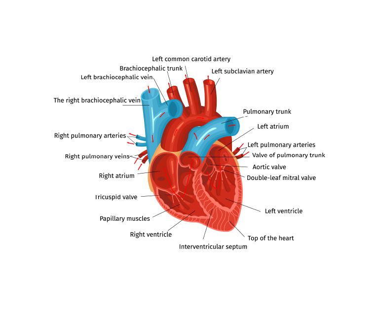
Heart Chambers
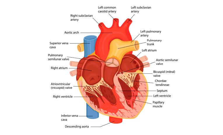
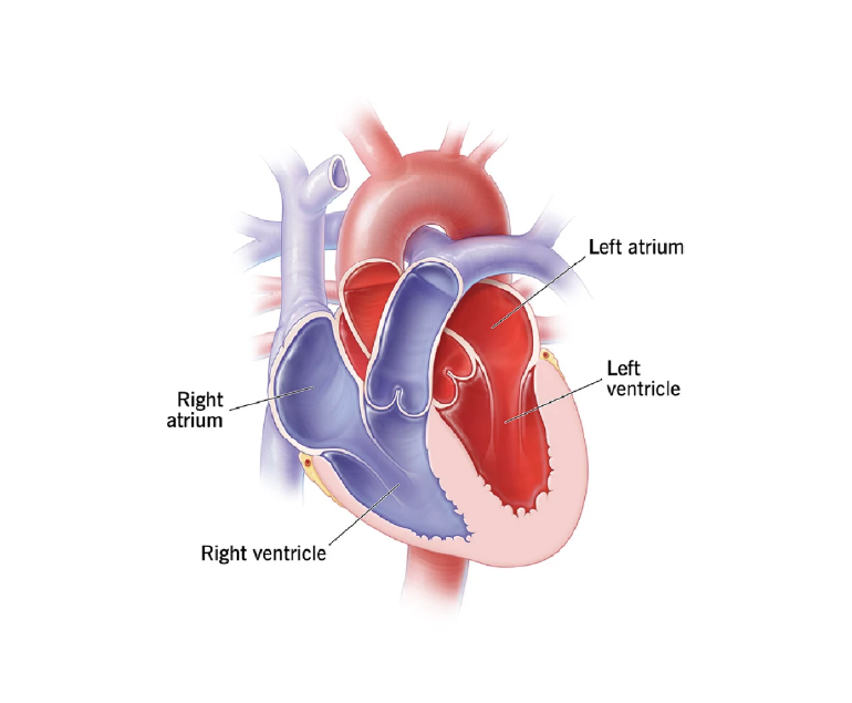
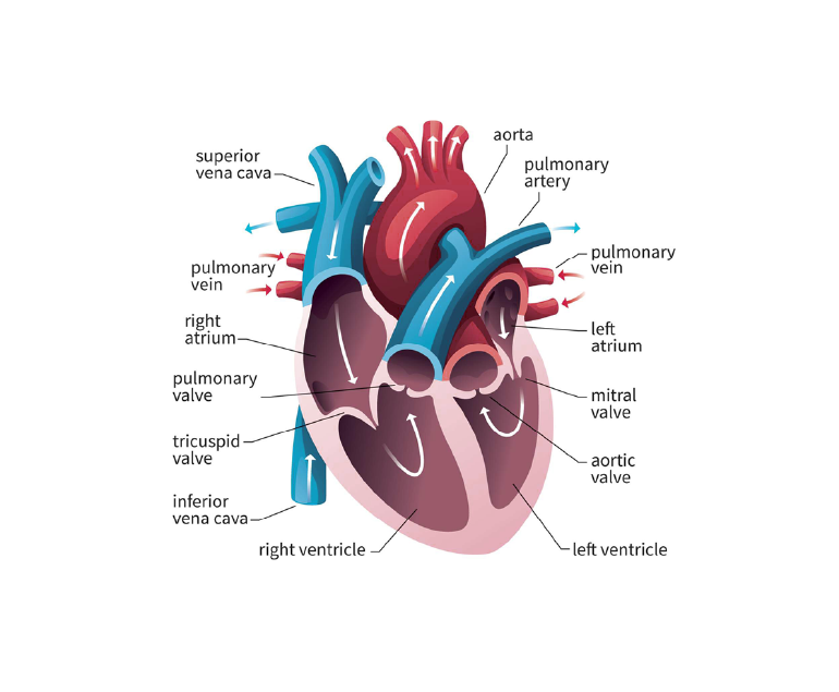
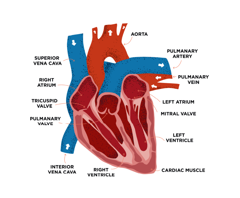
The heart is composed of four chambers: two atria and two ventricles. The atria are the upper chambers, while the ventricles are the lower chambers. The right atrium receives deoxygenated blood from the body through the superior and inferior vena cava. The blood then flows into the right ventricle, which pumps it into the lungs through the pulmonary artery for oxygenation.
Oxygenated blood from the lungs returns to the heart via the pulmonary veins and enters the left atrium. It is then pumped into the left ventricle, which propels it into the aorta, the main artery that carries oxygen-rich blood to the entire body.
Heart Valves
To ensure that blood flows in the correct direction, the heart is equipped with valves. There are two main types of valves: atrioventricular valves and semilunar valves. The atrioventricular valves, the tricuspid valve on the right side and the mitral valve on the left side, separate the atria from the ventricles.
They open to allow blood to flow from the atria to the ventricles and close to prevent backflow during contraction. The semilunar valves, including the pulmonic valve and the aortic valve, are located at the exit of the ventricles. They open to allow blood to be ejected from the heart and close to prevent blood from flowing back into the ventricles.
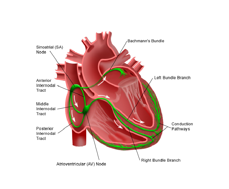
Heart Conduction System
The heart muscle itself, called the myocardium, is made up of specialized cardiac muscle cells. These cells have the remarkable ability to generate their electrical impulses, allowing the heart to beat independently of external nerve stimulation.
The electrical impulses start in the sinoatrial (SA) node, located in the right atrium. The impulses then spread through the atria, causing them to contract and push blood into the ventricles. The impulses are then conducted to the atrioventricular (AV) node, located in the septum between the atria.
From the AV node, the impulses travel down the ‘bundle of His’, a specialized electrical pathway, and its branches, which then stimulate the ventricles to contract, pushing blood out of the heart.
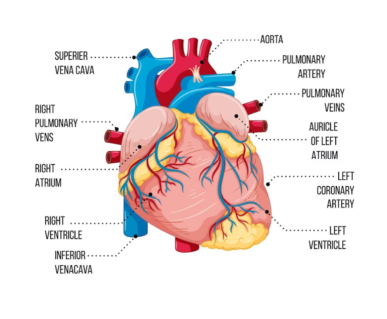
Blood Supply to the Heart
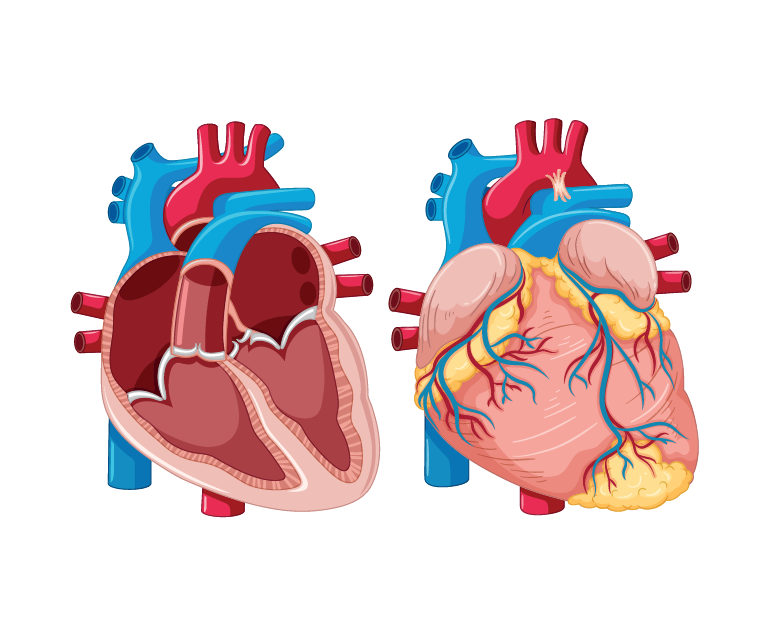
To meet the high metabolic demands of the heart muscle itself, a separate network of blood vessels called coronary arteries supplies oxygenated blood to the heart. The right coronary artery and the left coronary artery are the two main branches that arise from the aorta and encircle the heart.
These arteries give rise to smaller branches, known as coronary arterioles, which supply blood to the myocardium. The coronary veins collect the deoxygenated blood from the myocardium and drain it into the coronary sinus, which empties into the right atrium.
Importance of understanding the anatomy of the heart
Understanding the anatomy of the heart is crucial for diagnosing and treating various cardiac conditions. For example, a blockage in one or more of the coronary arteries can lead to reduced blood supply to the heart muscle, causing chest pain known as angina or even a heart attack.
Malfunctioning heart valves can result in conditions such as mitral valve prolapse or aortic stenosis, affecting the heart’s ability to pump blood efficiently. A thorough understanding of the heart’s anatomy allows healthcare professionals to identify and address these issues effectively.
Conclusion
The heart is a remarkable organ that plays a vital role in sustaining life. Its complex structure, with four chambers, valves, and its own blood supply, enables it to pump oxygenated blood to the entire body.
The heart’s anatomy and function are intricately interlinked, ensuring a continuous flow of oxygen and nutrients to every cell. Understanding the anatomy of the heart is crucial for appreciating how it functions and how it can be affected by various medical conditions.