Hypertrophic Cardiomyopathy

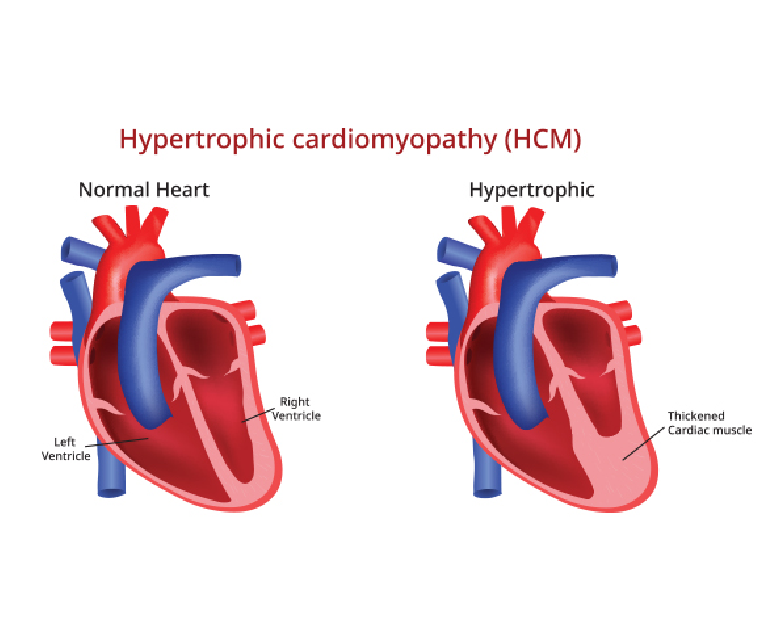
Hypertrophic cardiomyopathy (HOCM) is a complex and potentially life-threatening heart condition characterized by abnormal thickening (hypertrophy) of the heart muscle. It is one of the most common genetic heart disorders, affecting people of all ages and backgrounds.

Causes

Hypertrophic cardiomyopathy is primarily caused by mutations in genes responsible for the structure and function of the heart muscle. These genetic mutations lead to abnormal growth and arrangement of the cardiac muscle cells, resulting in the characteristic thickening of the heart muscle.

HOCM is usually inherited in an autosomal dominant pattern, meaning that a child has a 50% chance of inheriting the condition if one of the parents is affected. However, spontaneous gene mutations can also occur, leading to the development of HOCM in individuals without a family history of the condition.

Symptoms

The symptoms of hypertrophic cardiomyopathy can vary widely among individuals. Some may remain asymptomatic throughout their lives, while others may experience severe symptoms. Common symptoms include shortness of breath, chest pain, heart palpitations, fatigue, dizziness, fainting, and lightheadedness. These symptoms can worsen during physical exertion or strenuous activities and may be indicative of an increased risk of sudden cardiac arrest, especially in young athletes.

Diagnosis

The diagnosis of hypertrophic cardiomyopathy involves a comprehensive evaluation of the patient’s medical history, a physical examination, and a variety of diagnostic tests. These tests may include electrocardiography (ECG), echocardiography, cardiac MRI, exercise stress tests, and genetic testing. ECG can help identify abnormal electrical patterns in the heart, while echocardiography and cardiac MRI provide detailed images of the heart’s structure and function. Genetic testing is performed to identify specific gene mutations associated with HOCM.

Treatment

The management of hypertrophic cardiomyopathy focuses on relieving symptoms, preventing complications, and reducing the risk of sudden cardiac arrest. Treatment plans are tailored to individual patients based on their symptoms, risk factors, and disease progression. Medications such as beta-blockers, calcium channel blockers, and anti-arrhythmic drugs are commonly prescribed to control heart rate, rhythm, and blood pressure.

In some cases, surgical procedures like septal myectomy or alcohol septal ablation may be recommended to relieve obstruction of blood flow from the heart. Implantable cardioverter-defibrillators (ICDs) may also be used in high-risk patients to prevent sudden cardiac arrest.

Prognosis

The prognosis for individuals with hypertrophic cardiomyopathy varies depending on the severity of the disease, the presence of complications, and the effectiveness of treatment. Many people with HOCM lead normal lives with minimal symptoms and a low risk of complications. Regular monitoring, adherence to treatment plans, and lifestyle modifications, such as avoiding strenuous activities, can help improve outcomes and reduce the risk of complications.

Conclusion

Hypertrophic cardiomyopathy is a complex heart condition characterized by abnormal thickening of the heart muscle. It is primarily caused by genetic mutations and can present with a range of symptoms and complications. Early diagnosis, thorough evaluation, and personalized treatment plans are essential for managing HOCM effectively. With appropriate medical care and lifestyle modifications, individuals with hypertrophic cardiomyopathy can lead fulfilling lives while minimizing the risks associated with the condition.

Dilated Cardiomyopathy

Dilated cardiomyopathy (DCM) is a condition characterized by the dilation and weakening of the heart’s main pumping chamber, the left ventricle. This chronic and progressive disorder affects the heart muscle, leading to impaired cardiac function and potentially causing heart failure. DCM can also affect the right ventricle and both ventricles in some cases.

Causes

The exact causes of DCM are often unknown, but various factors can contribute to its development. These include genetic mutations, viral infections (such as viral myocarditis), exposure to toxins (for example alcohol), autoimmune diseases, and metabolic disorders. In many cases, DCM is considered idiopathic, meaning the cause is uncertain.

Symptoms

DCM can present with a range of symptoms, which may include fatigue, shortness of breath, swelling of the legs and ankles (edema), rapid or irregular heartbeat (arrhythmias), chest pain, dizziness, and fainting. These symptoms are often related to the heart’s inability to effectively pump blood and supply oxygen to the body’s tissues.

Diagnosis

To diagnose DCM, a comprehensive evaluation is required, which typically involves a thorough medical history review, physical examination, and various diagnostic tests. These tests may include electrocardiogram (ECG), echocardiogram (to assess heart structure and function), cardiac MRI or CT scan (for detailed imaging), blood tests (to rule out other conditions), and occasionally a heart biopsy.

Treatment

The management of DCM aims to improve symptoms, slow disease progression, and prevent complications. Treatment approaches may include:

  1. Medications: Several medications can be prescribed to manage symptoms, reduce the workload on the heart, control blood pressure, prevent blood clots, and treat arrhythmias. These may include beta-blockers, ACE inhibitors, diuretics, anticoagulants, and anti-arrhythmic drugs.
  • Lifestyle modifications: Patients need to make lifestyle changes like reducing salt intake, avoiding alcohol, stopping smoking, maintaining proper weight, and exercising as required.
  • Device therapy: In advanced cases or those with severe heart failure, implantable devices like pacemakers or defibrillators may be recommended to regulate the heart’s rhythm or deliver electrical shocks if life-threatening arrhythmias occur.
  • Cardiac rehabilitation: This supervised program combines exercise, education, and counseling to help patients recover and improve their overall cardiovascular health.
  • Surgical interventions: In some cases, surgical interventions may be necessary, such as coronary artery bypass grafting (CABG) to improve blood flow to the heart, or heart transplantation for end-stage heart failure.

Prognosis

The prognosis for individuals with DCM can vary widely depending on the underlying cause, severity of symptoms, and response to treatment. With appropriate management, lifestyle modifications, and adherence to medication, many patients can experience an improved quality of life and a slower disease progression. However, some individuals may develop more severe heart failure and require advanced therapies or heart transplantation. Regular follow-up with a cardiologist is essential to monitor the condition and adjust treatment as needed.

Conclusion

Dilated cardiomyopathy is a complex heart condition characterized by the enlargement and weakening of the heart muscle. While the exact causes are unclear in many cases, a combination of genetic, viral, autoimmune, and environmental factors can contribute to its development. Early diagnosis, appropriate medical management, lifestyle modifications, and sometimes surgical interventions are essential for managing the symptoms, slowing disease progression, and improving the prognosis for individuals with DCM.

Abdominal Aortic Aneurysm

Abdominal aortic aneurysm (AAA) is a potentially life-threatening condition characterized by the enlargement and weakening of the aorta—the main blood vessel that supplies oxygenated blood to the abdomen, pelvis, and lower extremities. AAA often develops slowly over time, and if left untreated, it can lead to a rupture, resulting in severe internal bleeding and potentially fatal consequences.

Causes

According to Dr. Ramji Mehrotra, India’s best heart surgeon, the exact cause of AAA is still unknown, but several risk factors have been identified. The primary risk factor is age, with individuals over 65 years being at higher risk. Other factors include smoking, high blood pressure, atherosclerosis (buildup of fatty deposits in the arteries), male gender, family history of AAA, and certain genetic disorders, such as Marfan syndrome and Ehlers-Danlos syndrome.

Symptoms

AAA often develops asymptomatically and is frequently detected incidentally during medical imaging tests conducted for unrelated conditions. However, as the aneurysm grows larger, it may cause symptoms such as:

  • Abdominal pain or discomfort: Dull, persistent pain or a pulsating sensation in the abdomen or back.
  • Swelling or a visible mass: A throbbing lump may be felt in the abdomen.
  • Rupture symptoms: Sudden and severe pain, dizziness, rapid heartbeat, low blood pressure, and loss of consciousness may occur if the aneurysm ruptures.

Diagnosis

AAA can be diagnosed through various imaging techniques, including:

  • Ultrasound: This non-invasive test uses sound waves to create images of the abdominal aorta, providing information about its size and structure.
  • Computed Tomography (CT) Scan: This imaging technique produces detailed cross-sectional images of the aorta, allowing for precise measurement and evaluation of the aneurysm.
  • Magnetic Resonance Imaging (MRI): MRI can provide additional information about the aorta and surrounding structures, especially in cases where CT scanning is contraindicated.

Treatment

The management of AAA depends on the size, growth rate, and overall health of the patient. Treatment options include:

  1. Watchful Waiting: Small aneurysms (less than 5.5 centimeters) that are not rapidly growing may be monitored regularly through imaging tests. Lifestyle modifications, such as quitting smoking and controlling blood pressure, are advised to reduce the risk of rupture.
  2. Surgery: For larger aneurysms or those growing rapidly, surgical intervention is often recommended. The two primary surgical procedures are open repair and endovascular aneurysm repair (EVAR).
  3. Open Repair: Involves surgically replacing the weakened section of the aorta with a synthetic graft.
  4. EVAR: A minimally invasive procedure where a stent graft is placed inside the aneurysm, providing support to the weakened vessel walls.

Conclusion

Dr. Ramji Mehrotra says that abdominal aortic aneurysm is a potentially life-threatening condition that requires careful monitoring and prompt management. Early diagnosis, regular screenings for at-risk individuals, and lifestyle modifications play a crucial role in preventing complications. It is important to consult with a doctor for accurate diagnosis, and appropriate treatment options.