Atherosclerosis

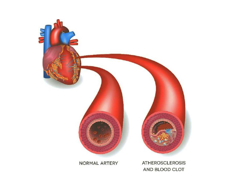
Atherosclerosis is a progressive condition in which the arteries become narrowed and hardened due to the buildup of plaque on their inner walls. Here is a more detailed explanation of the key aspects of atherosclerosis:

Plaque Formation: The process begins with damage to the inner lining (endothelium) of an artery. High blood pressure, smoking, high levels of cholesterol and triglycerides, and inflammation are some of the factors that can cause this damage. The endothelium’s damage causes the accumulation of substances like low-density lipoprotein (LDL) cholesterol and white blood cells at the site of injury.

Inflammatory Response: The immune system responds to the accumulation of these substances by initiating an inflammatory response. This causes the formation of fatty streaks, which are the early stages of plaque development.

Plaque Growth: Over time, the fatty streaks can progress into more complex plaques. These plaques consist of a core of cholesterol, calcium deposits, and other substances covered by a fibrous cap. As the plaque grows, it can protrude into the arterial lumen, narrowing the blood vessel.

Arterial Narrowing and Hardening: The accumulation of plaque leads to the narrowing of the arteries, reducing blood flow to organs and tissues. The plaque can also harden and calcify, making the arteries less elastic.

Reduced Blood Flow and Ischemia: As the arteries become progressively narrowed, the blood flow to vital organs and tissues decreases. This reduced blood flow can result in ischemia, which is an insufficient supply of oxygen and nutrients to the affected tissues.

Clinical Consequences: The consequences of atherosclerosis depend on the location of the affected arteries. If it occurs in the coronary arteries, it can lead to coronary artery disease and may result in angina (chest pain) or a heart attack. Atherosclerosis in the arteries supplying the brain can lead to cerebrovascular disease and increase the risk of stroke. In the peripheral arteries, it can cause peripheral artery disease, affecting the limbs and causing symptoms such as pain and difficulty walking.

Risk Factors and Prevention: Risk factors for atherosclerosis include high blood pressure, smoking, high cholesterol levels, diabetes, obesity, and a sedentary lifestyle. Prevention and management strategies involve adopting a healthy lifestyle, including regular exercise, a balanced diet, maintaining a healthy weight, managing blood pressure and cholesterol levels, and avoiding smoking.

Treatment approaches may include lifestyle modifications, medications to control risk factors, and, in some cases, invasive procedures such as angioplasty or bypass surgery to address severely blocked arteries. Early detection and management are crucial in preventing the progression of atherosclerosis and its associated complications.

Heart Transplantation Procedure

A heart transplant is a complex surgical procedure that involves replacing a diseased or failing heart with a healthy donor heart. Here’s an overview of the typical steps involved in a heart transplant procedure:

Patient Evaluation:

Before a heart transplant, a thorough evaluation of the patient’s medical history, current health status, and overall suitability for the procedure is conducted. This includes assessments of the patient’s heart function, other organ functions, and overall health.

Placement on the Transplant List:

If the patient is deemed a suitable candidate for a heart transplant, they are placed on a waiting list for a donor heart. The allocation of donor organs is based on factors such as blood type, body size, and medical urgency.

Waiting for a Donor:

Patients awaiting a heart transplant must often wait for a suitable donor heart. This waiting period can vary depending on factors such as the patient’s blood type, geographic location, and the availability of donor organs.

Donor Organ Retrieval:

Once a suitable donor heart becomes available, a surgical team is dispatched to retrieve the heart from the donor. The donor heart is carefully preserved and transported to the recipient’s hospital.

Recipient Surgery Preparation:

The recipient is prepared for surgery. This involves ensuring that the patient is in optimal condition for the transplant and ready to receive the donor heart.

Surgery:

The actual transplant surgery begins with the removal of the recipient’s diseased or failing heart. The donor heart is then implanted, and the surgical team connects the blood vessels and the heart’s chambers to ensure proper blood flow.

Recovery:

Following the surgery, the patient is closely monitored in the intensive care unit (ICU) for several days to weeks. This period is critical for assessing the success of the transplant and managing potential complications.

Post-Transplant Care:

After leaving the ICU, the patient continues to receive post-transplant care, including medications to prevent organ rejection. Regular follow-up appointments with the transplant team are crucial for long-term monitoring and adjustments to the treatment plan.

It’s important to note that heart transplant surgery is a major undertaking with associated risks and complications. The success of the procedure depends on various factors, including the overall health of the patient, the compatibility of the donor organ, and the expertise of the surgical and medical teams. Patients who undergo heart transplants often require lifelong medical care and immunosuppressive medications to prevent rejection of the transplanted organ.

Heart Health Precautions during the Winter

Maintaining heart health during the winter is important, as colder temperatures can affect the cardiovascular system. Here are some precautions to help promote heart health during the winter:

Stay Active:

Exercise regularly, even during the winter. Consider outdoor activities like walking, swimming. If you exercise outdoors, dress in layers to stay warm, and be cautious of icy conditions.

Keep Warm:

Dress in layers to stay warm, especially if you’re going outside. Cold temperatures can cause blood vessels to constrict, raising blood pressure. Wearing warm clothing helps to maintain body heat and prevent this. Avoid sudden change of temperature.

Stay Hydrated:

It’s easy to forget to drink enough water during the colder months, but staying hydrated is crucial for heart health. Dry winter air can also contribute to dehydration.

Maintain a Healthy Diet:

Eat a balanced and heart-healthy diet. Include plenty of fruits, vegetables, whole grains, and lean proteins. Limit the intake of saturated and fats, sodium, and added sugars.

Manage Stress:

Practice stress-reduction techniques, as stress can impact heart health. Consider activities like meditation, deep breathing exercises, or yoga to manage stress levels.

Stay Warm Indoors:

Cold indoor temperatures can also affect your cardiovascular system. Keep your home at a comfortable temperature, and use blankets if needed. Avoid prolonged exposure to cold indoor environments.

Take precautions when shoveling snow.

If you need to shovel snow, take breaks and lift small amounts at a time to avoid putting too much strain on your heart. If you have cardiovascular issues, consult your doctor before shoveling snow.

Keep up with medications:

If you’re on medications for heart-related conditions, continue taking them as prescribed. If you have concerns or experience any side effects, consult your healthcare provider.

Stay Informed:

Be aware of weather conditions, especially extreme cold or winter storms. Plan accordingly and avoid unnecessary exposure to severe/extreme weather conditions.

Get Vaccinated:

Influenza (flu) and other respiratory infections are more common during the winter. Consider getting vaccinated to reduce the risk of these illnesses, which can put extra stress on the heart.

Regular Check-ups:

Schedule regular check-ups with your healthcare provider. This is especially important if you have pre-existing cardiovascular conditions. Regular monitoring can help catch potential issues early.

Always consult with your healthcare provider before making significant changes to your lifestyle or if you have specific concerns about your heart health, especially during the winter months. They can provide personalized advice based on your individual health status.( Personalized advice can be given according to your specific health status.)

Always consult with your healthcare provider before making significant changes to your lifestyle or if you have specific concerns about your heart health, especially during the winter months. They can provide personalized advice based on your individual health status.( Personalized advice can be given according to your specific health status.)

Schedule regular check-ups with your healthcare provider. This is especially important if you have pre-existing cardiovascular conditions. Regular monitoring can help catch potential issues early.

Air Pollution & Heart Disease

Air pollution is considered to be a silent killer that affects our daily lives. From the exhaust fumes of vehicles to industrial emissions and even natural sources like dust storms, the air we breathe is often contaminated with many harmful particles and gases. While we often associate respiratory problems with air pollution, its impact on heart health is equally concerning.

Effect of Air Pollution on the Heart

Particulate matter (PM) and gaseous pollutants are known to cause great harm to the cardiovascular system. Fine particulate matter, known as PM2.5, consists of tiny particles less than 2.5 micrometers in diameter. These minuscule particles can penetrate deep into the lungs and even enter the bloodstream. Once the particles enter the bloodstream, they can trigger a cascade of reactions that contribute to the development and progression of heart disease.

  1. Inflammation: PM2.5 and other pollutants can induce inflammation in the arteries. Inflammatory responses can lead to the buildup of plaque in the arteries, a condition known as atherosclerosis. Atherosclerosis narrows the arteries and restricts blood flow to the heart, increasing the risk of heart attacks and strokes.
  2. Blood pressure: Air pollution has been linked to increased blood pressure. Over time, elevated blood pressure can strain the heart and lead to conditions like hypertension and hypertensive heart disease. These conditions make the heart work harder and can result in heart failure.
  3. Irregular heart rhythms: Some studies have suggested that air pollution may contribute to irregular heart rhythms, known as arrhythmias. These irregular heartbeats can be life-threatening, especially in individuals with pre-existing heart conditions.
  4. Heart attacks: The risk of heart attacks, or myocardial infarctions, is significantly higher in areas with high levels of air pollution. Pollutants can trigger the rupture of existing plaques in the arteries, leading to blood clots that can block blood flow to the heart muscle.
  5. Heart failure: Air pollution can aggravate existing heart conditions, such as heart failure, by increasing the workload on the heart. Heart failure occurs when the heart cannot pump blood effectively, leading to a buildup of fluid in the lungs and other tissues.

Who Is at Risk?

Leading cardiovascular surgeon Dr. Ramji Mehrotra cites the example of the high levels of air pollution in New Delhi and is of the opinion that while air pollution poses a risk to everyone, certain groups are more vulnerable to its adverse effects on the heart. These include:

  1. Elderly individuals: Older adults are more likely to have pre-existing heart conditions, making them more susceptible to the harmful effects of air pollution.
  2. Children: Developing hearts in children are more sensitive to environmental stressors, including air pollution, which can have long-term implications for their heart health.
  3. People with pre-existing heart conditions: Those with conditions like coronary artery disease, hypertension, and heart failure are at greater risk of experiencing adverse cardiovascular effects from air pollution.
  4. Individuals with low socioeconomic status: People in lower-income communities often face higher exposure to air pollution due to factors like living closer to industrial areas and having limited access to healthcare resources.

Reducing the Risk

Considering the link between air pollution and heart disease, it is crucial to take steps to reduce the risk. Here are some strategies to protect your heart:

  1. Stay informed: Monitor local air quality and avoid outdoor activities on days when air pollution levels are high.
  2. Use air purifiers: Indoor air pollution can also be a concern, so using air purifiers at home can help reduce exposure to harmful particles.
  3. Reduce vehicle emissions: Carpooling, using public transportation, and driving fuel-efficient vehicles can help reduce the emissions that contribute to outdoor air pollution.
  4. Plant trees and vegetation: Greenery can act as natural air filters and improve local air quality. Supporting and participating in urban reforestation efforts can make a difference.
  5. Advocate for clean energy: Supporting policies and initiatives that promote clean energy sources and reduce emissions is essential for long-term improvement in air quality.
  6. Lead a heart-healthy lifestyle: A heart-healthy diet, regular exercise, and stress management can help mitigate the effects of air pollution on heart health.
  7. Avoid Smoking and tobacco use.

Conclusion

Dr. Mehrotra is of the opinion that Air Pollution is not only a threat to our respiratory health but also a silent contributor to heart disease. The tiny particles and harmful gases in polluted air can have profound and lasting effects on the cardiovascular system, increasing the risk of heart attacks, high blood pressure, and other heart-related conditions. Protecting your heart from air pollution involves a combination of individual actions, community efforts, and policy changes. By taking these steps, we can breathe easier and ensure a healthier future for our hearts.

Pediatric Heart Infections

Pediatric heart infections, although relatively rare, can have serious and potentially life-threatening consequences for children. These infections affect the heart muscle, valves, or the inner lining of the heart chambers and are often caused by bacteria, viruses, or other microorganisms. Early detection, timely treatment, and a good understanding of the risks associated with pediatric heart infections are crucial for the well-being of affected children. In this blog, Dr. Ramji Mehrotra, India’s leading pediatric cardiac surgeon, elaborates upon the causes, symptoms, diagnosis, and treatment options for these infections.

Causes of Pediatric Heart Infections

  • Bacterial Endocarditis: This is one of the most common types of heart infections in children. It occurs when bacteria enter the bloodstream and attach to the heart valves or lining of the heart. Certain pre-existing heart conditions, congenital heart defects, or the use of intravenous (IV) lines and catheters can increase the risk of bacterial endocarditis.
  • Viral Myocarditis: Myocarditis is an inflammation of the heart muscle, often caused by viral infections such as enteroviruses or adenoviruses. While viral myocarditis is less common in children, it can lead to significant cardiac issues if left untreated.

Symptoms

The symptoms of pediatric heart infections can vary depending on the type and severity of the infection. Common symptoms include:

  • Fever: A persistent fever that doesn’t respond to regular treatments.
  • Fatigue: Unexplained tiredness or weakness.
  • Chest Pain: Discomfort especially during physical activity.
  • Rapid Breathing: Increased breathing rate, especially in infants.
  • Paleness: Pale or bluish skin, lips, or fingertips.
  • Joint Pain: Joint pain or swelling.
  • Swelling: Swelling in the legs and abdomen
  • Abnormal Heartbeat: Irregular heartbeat, known as arrhythmia.
  • Poor Feeding: Difficulty feeding in infants.
  • Weight Loss: Unintentional weight loss.
  • Irritability: Increased fussiness or irritability.

Diagnosis

Diagnosing pediatric heart infections can be challenging because the symptoms can mimic those of other common childhood illnesses. Physicians typically use a combination of the following diagnostic tools:

  1. Physical Examination: A thorough physical examination to check for symptoms and signs of infection.
  2. Blood Tests: Blood tests to check for infection markers and the presence of microorganisms.
  3. Electrocardiogram (ECG or EKG): A test that records the heart’s electrical activity to identify irregularities in the heartbeat.
  4. Echocardiogram: An ultrasound of the heart to visualize its structure and function.
  5. Chest X-rays: X-rays to assess the heart and lungs.
  6. MRI or CT Scan: These imaging tests may be used to provide detailed images of the heart.

Treatment

Treatment of pediatric heart infections typically involves a combination of medical therapies and, in some cases, surgical intervention, depending on the type and severity of the infection. Common treatment options include:

  1. Antibiotics: Bacterial infections are treated with antibiotics.
  2. Antiviral Medications: Viral myocarditis may be treated with antiviral drugs.
  3. Anti-inflammatory Medications: Medications like corticosteroids may be used to reduce inflammation in the heart.
  4. Surgery: In severe cases or when complications arise, surgical intervention may be necessary. This can include repairing damaged heart valves or removing infected tissue.
  5. Supportive Care: Children with heart infections may require hospitalization for supportive care, such as intravenous fluids, pain management, and monitoring of vital signs.

Conclusion

According to Dr. Ramji Mehrotra, pediatric heart infections are a serious concern, but with early detection and appropriate treatment, the prognosis can be favourable. Parents and healthcare providers must be vigilant about recognizing the symptoms and risk factors associated with these infections. Additionally, maintaining good hygiene practices, ensuring proper dental care, and managing pre-existing heart conditions can help reduce the risk of pediatric heart infections. Overall, a proactive approach to heart health can make a significant difference in the well-being of children.

Minimally Invasive Direct Coronary Artery Bypass Grafting

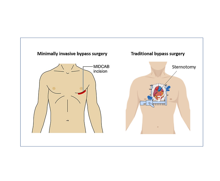
Minimally Invasive Direct Coronary Artery Bypass Grafting (MIDCAB) is a less invasive surgical procedure for treating coronary artery disease (CAD). Unlike traditional coronary artery bypass grafting (CABG), which requires a median sternotomy and the use of a heart-lung machine, MIDCAB is performed through a small incision on the left side of the chest, offering several advantages.

Procedure Highlights

  1. Small Incision: MIDCAB involves a small incision between the ribs, eliminating the need for a sternotomy. This results in less chest trauma and reduced postoperative pain.
  2. On-Beating Heart: Unlike traditional CABG, which temporarily stops the heart with a heart-lung machine, MIDCAB is performed on a beating heart. This reduces the risk of complications associated with bypassing the heart’s normal function.
  3. Shorter Recovery: Patients undergoing MIDCAB experience a shorter hospital stay and a faster return to their regular activities, making it an appealing option for those seeking quicker recovery.
  4. Reduced Scarring: The small incision in MIDCAB results in less visible scarring, providing cosmetic benefits for some patients.

Patient Eligibility

MIDCAB is suitable for patients with single or double-vessel disease, meaning they have blockages in one or two coronary arteries. It is not appropriate for complex multi-vessel disease requiring multiple grafts. Favorable patient anatomy and good overall health are essential for candidacy.

Procedure Steps

  1. Incision: A small incision is made between the ribs on the left side of the chest to access the target coronary artery.
  2. Graft Placement: A healthy blood vessel, often the internal mammary artery or radial artery, is harvested and prepared for grafting. It is then attached to the blocked coronary artery, creating a new pathway for blood flow.
  3. Heart Monitoring: Specialized equipment is used to monitor the heart throughout the procedure to ensure it continues to beat.
  4. Closure: After successfully placing the graft, the incision is closed, and the chest is sutured. No sternotomy is required.

Benefits and Considerations

Advantages:

  1. Less Invasive: MIDCAB results in reduced chest trauma, postoperative pain, and discomfort.
  2. Quicker Recovery: Patients have a faster recovery and shorter hospital stay.
  3. Cosmetic Appeal: The small incision leads to less visible scarring.

Considerations:

  1. Limited Applicability: MIDCAB is suitable only for patients with single or double-vessel disease.
  2. Long-Term Efficacy: Long-term outcomes compared to traditional CABG are still under investigation.
  3. Operator Skill: MIDCAB requires a highly skilled and experienced surgeon.
  4. Patient Selection: Careful patient evaluation is crucial to ensure the best outcomes.

Summary

Minimally Invasive Direct Coronary Artery Bypass Grafting (MIDCAB) is an attractive option for many patients with CAD due to its less invasive nature, faster recovery, and improved cosmetic results. However, patient suitability and careful evaluation by doctors are key factors in determining the most appropriate treatment approach.

Bicuspid Aortic Valve

The human heart is a remarkable organ that is responsible for the continuous flow of oxygen-rich blood throughout the body. Among its critical components is the aortic valve, a gateway between the left ventricle and the aorta, which ensures that blood is propelled in the right direction. Typically, the aortic valve has three parts, or leaflets.

A bicuspid aortic valve, or BAV, is a congenital heart condition characterized by the presence of only two leaflets in the aortic valve. This condition affects approximately 1-2% of the population, making it one of the most common congenital heart abnormalities. While it may seem like a minor anatomical deviation, BAV can have significant implications for a person’s heart health.

Anatomy

To understand the significance of BAV, it is essential to know a bit about the aortic valve’s structure. In a healthy heart, the aortic valve is comprised of three thin, flexible leaflets, which open and close with each heartbeat. This rhythmic movement ensures that oxygenated blood flows from the left ventricle into the aorta and onward to the rest of the body.

In contrast, individuals with a bicuspid aortic valve have only two leaflets, which can be fused together to varying degrees. This fusion creates a valve that is less efficient at regulating blood flow. As a result, the valve may become stenotic (narrowed) or insufficient (leaky), causing a range of problems.

Complications and Symptoms

BAV can lead to several complications, with aortic stenosis and aortic regurgitation being the most common. Aortic stenosis occurs when the valve opening narrows, restricting blood flow. Aortic regurgitation, on the other hand, results from a leaky valve, causing blood to flow backward into the left ventricle.

Individuals with BAV may experience symptoms such as chest pain, shortness of breath, palpitations, and fatigue. If left untreated, BAV can lead to serious heart conditions, including heart failure, arrhythmias, and aortic aneurysms. Therefore, early detection and management of BAV are crucial.

Diagnosis and Treatment

Diagnosing BAV typically involves echocardiography, a non-invasive imaging technique that provides detailed pictures of the heart and its valves. Once diagnosed, the treatment plan depends on the severity of the condition. Some individuals with BAV may require no treatment if the valve functions adequately. However, those with significant stenosis or regurgitation may need medications, valve repair, or replacement surgery.

In recent years, transcatheter aortic valve replacement (TAVR) has emerged as a less invasive alternative to open-heart surgery for some BAV patients. TAVR involves the insertion of a collapsible valve through a catheter, which is then expanded inside the aorta to replace the dysfunctional valve.

Conclusion

A bicuspid aortic valve is a unique heart condition that is relatively common but can have a significant impact on heart health. Advances in diagnosis and treatment have improved the outlook for people with BAV, highlighting the importance of ongoing research and medical innovation in the field of cardiology.

Cardiovascular Disease and Pregnancy

Pregnancy is a transformative and joyous experience in a woman’s life, but it also places significant physiological demands on the body. For women with pre-existing cardiovascular disease, pregnancy can present unique challenges and risks. Cardiovascular disease encompasses a range of conditions that affect the heart and blood vessels, including hypertension, congenital heart defects, valvular heart disease, and cardiomyopathy. Managing cardiovascular disease during pregnancy requires careful planning, monitoring, and collaboration between obstetricians and cardiologists to ensure the health of both the mother and the developing fetus.

Risk Factors and Preconception Care

Women with pre-existing cardiovascular disease who plan to become pregnant should receive specialized preconception care. This involves a thorough assessment of their cardiovascular health, including a review of their medical history, current medications, and cardiac function. Understanding the specific type and severity of cardiovascular disease is essential for risk assessment and management planning.

Risks for Mother

During pregnancy, the body undergoes significant changes to support fetal growth. Blood volume increases, heart rate rises, and the cardiovascular system works harder to meet the body’s increased oxygen and nutrient demands. For women with cardiovascular disease, these changes can aggravate their condition and lead to complications such as heart failure, arrhythmias, and thromboembolic events. Hypertension, in particular, can become more challenging to manage during pregnancy and is a significant risk factor for complications.

Risks for Fetus

The developing fetus relies on a well-functioning maternal cardiovascular system for oxygen and nutrient supply. Maternal cardiovascular disease can compromise blood flow to the placenta, potentially resulting in fetal growth restriction, preterm birth, or stillbirth. Additionally, certain medications used to manage cardiovascular conditions may have teratogenic effects on the developing fetus, highlighting the importance of careful medication management during pregnancy.

Management and Monitoring

Managing cardiovascular disease during pregnancy requires a multidisciplinary approach involving obstetricians, cardiologists, and other specialists as needed. Individualized treatment plans are crucial, taking into account the specific cardiovascular condition, its severity, and the patient’s overall health. Medications may need to be adjusted or changed to minimize potential harm to the fetus while ensuring maternal well-being.

Frequent monitoring is essential throughout pregnancy. This includes regular check-ups to assess blood pressure, cardiac function, and fetal growth. Advanced imaging techniques like echocardiography and Doppler ultrasound can provide valuable insights into maternal and fetal well-being.

Delivery Planning

The mode of delivery should also be carefully planned. In many cases, vaginal delivery is safe and appropriate, but a caesarean section may be recommended if there are concerns about the mother’s cardiovascular stability or the fetus’s well-being. The timing of delivery may also be adjusted to optimize outcomes for both mother and baby.

Postpartum Care

The challenges of managing cardiovascular disease do not end with delivery. The postpartum period is a critical time, as the body undergoes significant physiological changes once again. Close monitoring should continue, and adjustments to medications and treatment plans may be necessary.

Conclusion

Pregnancy with pre-existing cardiovascular disease requires meticulous planning, close monitoring, and a collaborative healthcare team. While it can be associated with increased risks for both the mother and the fetus, with proper management and care, many women with cardiovascular disease can have successful pregnancies and healthy babies. Education, counseling, and ongoing support are vital for empowering women with cardiovascular disease to make informed decisions about family planning and to navigate the complex journey of pregnancy with confidence.

Anxiety and Heart Disease

Anxiety is often considered as an emotional response to stress and has a profound impact on our physical well-being. While it is widely known that anxiety can take a toll on mental health, its intricate connection with heart disease is often underestimated. In this blog, we analyze the link between anxiety and heart disease, exploring how these seemingly unrelated facets of health are closely interlinked.

Basics of Anxiety

Anxiety is a normal response to stress, which is a built-in mechanism that helps human beings to navigate challenging situations. However, when anxiety becomes chronic or overwhelming, it can turn into a debilitating condition. Generalized Anxiety Disorder (GAD), Panic Disorder, and Social Anxiety Disorder are few of the anxiety-related conditions that affect millions of people daily.

The symptoms of anxiety can range from excessive worry and restlessness to physical manifestations like increased heart rate, muscle tension, and shortness of breath.

Connection between Anxiety and Heart Disease

Anxiety and heart disease are connected both biologically and psychologically. When we experience anxiety, our body goes into “fight or flight” mode. This response triggers the release of stress hormones like cortisol and adrenaline. While this response is essential in moments of acute danger, prolonged activation can negatively affect our cardiovascular system.

One of the immediate effects of stress hormones is an increase in heart rate and blood pressure. Over time, this can lead to a condition known as hypertension, or high blood pressure. Hypertension is a significant risk factor for heart disease. It places added strain on the heart, making it work harder and increasing the risk of various heart conditions, including coronary artery disease and heart attacks.

Moreover, chronic anxiety can lead to unhealthy lifestyle choices that further increase the risk of heart disease. Individuals with anxiety may be more likely to engage in smoking, excessive alcohol consumption, or overeating as a means of coping with their symptoms. These behaviors are all associated with an increased risk of heart disease.

Anxiety and heart disease also share a psychological connection. Living with anxiety can be very stressful, and chronic stress is detrimental to heart health. Prolonged stress can lead to unhealthy coping mechanisms, including emotional eating, lack of physical activity, obesity and social isolation.

Additionally, severe anxiety can also lead to depression. Depressive symptoms can contribute to a sedentary lifestyle and poor dietary choices, both of which are risk factors for heart disease. Moreover, individuals with anxiety and depression may have a reduced motivation to adhere to necessary heart-healthy behaviors, such as taking prescribed medications or attending medical appointments.

Prevention and Management

Some strategies that can be adopted for preventing and managing this dual threat to your health include:

  1. Stress Management: Learning effective stress management techniques, such as mindfulness, meditation, or deep breathing exercises, can help reduce the impact of anxiety on your cardiovascular system.
  2. Regular Physical Activity: Engaging in regular exercise is not only beneficial for your heart but can also help alleviate anxiety symptoms. Exercise releases endorphins, which are natural mood lifters.
  3. Healthy Diet: A balanced diet rich in fruits, vegetables, whole grains, and lean proteins can improve both your mental and heart health. Avoid excessive caffeine and sugar, as they can increase the levels of anxiety.
  4. Seek Professional Help: If anxiety is severely impacting your life, consider seeking professional help. Therapy, medication, or a combination of both can be effective in managing anxiety disorders.
  5. Medication Management: If you have existing heart conditions and are experiencing anxiety, discuss your symptoms with your doctors. They can prescribe a personalized treatment approach that considers both your heart health and anxiety.

Conclusion

The link between anxiety and heart disease highlights the importance of a holistic approach to health. Treating anxiety isn’t just about improving mental well-being; it is also about safeguarding your heart. By recognizing this connection and taking proactive steps to manage anxiety, you can reduce your risk of heart disease and live a healthier, happier life.

The link between anxiety and heart disease highlights the importance of a holistic approach to health. Treating anxiety isn’t just about improving mental well-being; it is also about safeguarding your heart. By recognizing this connection and taking proactive steps to manage anxiety, you can reduce your risk of heart disease and live a healthier, happier life.

Congenital Heart Disease in Children

Congenital heart disease (CHD) is a group of structural heart defects present at birth, affecting millions of children worldwide. These anomalies range from minor issues that may require minimal intervention to severe, life-threatening conditions. Understanding CHD, its causes, diagnosis, treatment, and the emotional toll it takes on families is crucial for providing comprehensive care and support.

Causes and Types of CHD

CHD originates during fetal development when the heart does not form correctly. While the exact causes remain unknown in many cases, genetic factors, maternal illnesses, and environmental factors may contribute. There are various types of CHD, classified into two main categories: cyanotic and acyanotic.

  • Cyanotic CHD: These defects cause decreased oxygen levels in the blood, resulting in bluish skin and lips. Examples include Tetralogy of Fallot and Transposition of the Great Arteries.
  • Acyanotic CHD: These defects do not lead to bluish discoloration but can still be serious. Examples include Ventricular Septal Defect (VSD) and Atrial Septal Defect (ASD).

Diagnosis and Screening

CHD is typically diagnosed during pregnancy through routine ultrasounds or shortly after birth when a newborn is screened for congenital anomalies. Early detection is crucial for prompt intervention and better outcomes.

Treatment and Management

The treatment approach for CHD varies depending on the type and severity of the defect. Some children may only require periodic monitoring, while others need surgical or interventional procedures. Advances in medical technology and surgical techniques have greatly improved the outlook for children with CHD.

  1. Medication: Some CHD cases can be managed with medication to control symptoms and improve heart function. For example, diuretics may help remove excess fluid buildup, while beta-blockers can regulate heart rate.
  2. Surgery: Surgical intervention is often necessary for complex CHD cases. Surgeons can repair heart defects, replace damaged valves, or even perform heart transplants when required.
  3. Interventional Procedures: Minimally invasive procedures like catheterization can be used to treat certain CHD cases. These procedures involve threading a catheter through blood vessels to repair or correct heart defects.

Psychosocial Impact

CHD not only affects the child but also places a significant emotional and financial burden on families. Parents often experience feelings of guilt, anxiety, and stress. Siblings may feel neglected, and the child with CHD may struggle with self-esteem and body image issues as they grow.

Supportive care, counseling, and access to support groups can help families navigate these challenges. A multidisciplinary approach involving pediatricians, cardiologists, social workers, and psychologists is essential to address both the medical and emotional aspects of CHD.

Long-term Outlook

Advancements in medical science have led to a brighter outlook for children with CHD. Many go on to lead healthy lives with appropriate care and monitoring. However, long-term follow-up is critical to identify and address any potential complications as they grow.

Conclusion

Congenital heart disease in children is a complex and challenging condition that requires comprehensive care and support. Early diagnosis and access to specialized medical care are vital for improving outcomes. Equally important is the emotional and psychosocial support provided to families as they navigate the journey of caring for a child with CHD. With continued research and advancements in medical technology, the future looks promising for children born with these heart defects, offering hope for healthier and happier lives.

Posts navigation