Ross Procedure

The Ross procedure is a surgical procedure used to treat aortic valve disease, which is a condition in which the valve that controls blood flow from the heart to the aorta becomes narrowed or leaks. This procedure involves replacing the damaged aortic valve with the patient’s own pulmonary valve, and then replacing the pulmonary valve with a donated or artificial valve.

The Ross procedure is named after its creator, Donald Ross, a Scottish cardiac surgeon who developed the technique in the 1960s. Since then, the procedure has become a popular option for treating aortic valve disease in certain patients.

Advantages of Ross Procedure

The Ross procedure is typically recommended for younger patients who have aortic valve disease, as it has several advantages over other types of valve replacement surgery. One of the main advantages is that it avoids the need for lifelong anticoagulation therapy, which is required for patients who receive a mechanical valve replacement. Anticoagulation therapy is a type of medication that helps prevent blood clots, but it can also increase the risk of bleeding and other complications.

Another advantage of the Ross procedure is that it provides a better quality of life for patients. The pulmonary valve is better suited to the low-pressure environment of the aortic position than artificial valves, which can often lead to complications such as valve dysfunction or blood clots.

Procedure

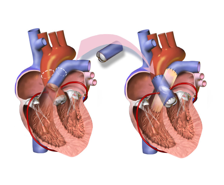
The Ross procedure is performed under general anaesthesia and typically takes many hours to complete. During the procedure, the surgeon will make an incision in the chest and access the heart. The patient’s pulmonary valve will then be removed and used to replace the damaged aortic valve. A new pulmonary valve, either from a donated cadaver or an artificial valve, will then be used to replace the removed pulmonary valve.

After the procedure, patients may need to spend several days in the hospital recovering. They may experience some discomfort or pain, and will need to take medication to manage pain and prevent infection. Patients will also need to gradually increase their physical activity and follow a prescribed rehabilitation program to help them recover.

Suitability

While the Ross procedure has many benefits, it is not suitable for all patients with aortic valve disease. Patients who have other heart conditions, such as coronary artery disease or an enlarged heart, may not be good candidates for the procedure. Additionally, patients who have already had a previous valve replacement or who have other medical conditions that could complicate surgery may not be good candidates for the procedure.

Conclusion

Ross procedure is a surgical procedure used to treat aortic valve disease in certain patients. It involves replacing the damaged aortic valve with the patient’s own pulmonary valve, and then replacing the pulmonary valve with a donated or artificial valve. If you are considering the Ross procedure, it is important to discuss the risks and benefits with your doctor to determine if it is the right option for you.

What is Peripheral Artery Disease?

Peripheral artery disease (PAD) is a circulatory condition that affects the peripheral arteries, which are the blood vessels that carry blood from the heart to the arms, legs, and other parts of the body. PAD occurs when these arteries become narrow or blocked, reducing blood flow to the affected body parts. The most common symptoms of PAD include pain, numbness, and cramping in the legs or feet, especially during physical activity.

How is PAD caused?

PAD is caused by atherosclerosis, a condition in which fatty deposits, or plaques, build up inside the arteries, narrowing the blood vessels and restricting blood flow. Atherosclerosis can affect any artery in the body, but it is most commonly found in the peripheral arteries of the legs and feet. Other risk factors for PAD include smoking, high blood pressure, high cholesterol, diabetes, obesity, and a family history of the disease.

PAD can be a serious condition, and if left untreated, it can lead to complications such as foot ulcers, gangrene, and amputation. However, there are a number of treatments available for PAD, and early diagnosis and treatment can help prevent these complications and improve quality of life.

Treatment

The first step in treating PAD is to make lifestyle changes that can help improve blood flow and reduce the risk of further narrowing of the arteries. These may include quitting smoking, losing weight, exercising regularly, and following a healthy diet.

Medications may also be prescribed to help manage symptoms and reduce the risk of complications. Some of the most commonly used medications for PAD include antiplatelet agents, such as aspirin or clopidogrel, which can help prevent blood clots from forming in the narrowed arteries, and statins, which can help lower cholesterol levels and reduce the risk of further plaque build-up.

In more severe cases of PAD, surgical or minimally invasive procedures may be necessary to restore blood flow to the affected areas. These procedures may include angioplasty, in which a small balloon is inserted into the narrowed artery and inflated to widen the artery and improve blood flow, or stenting, in which a small metal mesh tube is placed in the artery to hold it open.

In some cases, bypass surgery may be necessary, in which a healthy blood vessel is taken from another part of the body and used to bypass the blocked artery, allowing blood to flow freely to the affected area.

If left untreated, PAD can lead to serious complications, including foot ulcers, gangrene, and amputation. However, with proper diagnosis and treatment, the prognosis for PAD is generally good. By making lifestyle changes, taking medication as prescribed, and undergoing any necessary procedures, most people with PAD can improve their symptoms, reduce their risk of complications, and maintain their mobility and quality of life.

Conclusion

PAD is a common circulatory condition that can cause pain, numbness, and cramping in the legs and feet, especially during physical activity. It is caused by atherosclerosis, and risk factors include smoking, high blood pressure, high cholesterol, diabetes, obesity, and a family history of the disease. Treatment for PAD may include lifestyle changes, medications, or surgical procedures, depending on the severity of the condition. Early diagnosis and treatment can help prevent complications and improve quality of life for people with PAD.

Truncus Arteriosus

Truncus arteriosus is a rare congenital heart defect that occurs in approximately 1 in 10,000 live births. It is a serious condition that requires prompt medical attention and intervention to prevent potentially life-threatening complications.

What is Truncus arteriosus?

In a normal heart, the blood is pumped from the right side of the heart to the lungs, where it is oxygenated, and then returned to the left side of the heart, where it is pumped out to the rest of the body. This process is facilitated by two separate arteries, the pulmonary artery, which carries blood to the lungs, and the aorta, which carries blood to the rest of the body.

However, in the case of truncus arteriosus, the pulmonary artery and the aorta fail to separate during foetal development, resulting in a single large vessel that arises from both the right and left ventricles. This vessel is known as the truncus arteriosus. Additionally, there is usually a hole in the wall that separates the ventricles, called a ventricular septal defect (VSD).

As a result of this defect, oxygen-rich and oxygen-poor blood mix together in the truncus arteriosus before being pumped out to the body. This results in a lack of oxygen to the body’s tissues, leading to symptoms such as fatigue, shortness of breath, and poor growth.

Truncus arteriosus can also lead to more serious complications such as heart failure, pulmonary hypertension, and developmental delays. The severity of the condition depends on the size of the VSD and the degree of mixing of blood within the truncus arteriosus.

Diagnosis & Treatment

Truncus arteriosus is typically diagnosed soon after birth or during infancy, with symptoms such as a bluish tint to the skin (cyanosis), rapid breathing, and poor feeding. Diagnostic tests such as echocardiography, cardiac catheterization, and magnetic resonance imaging (MRI) may be performed to confirm the diagnosis and evaluate the severity of the defect.

Treatment for truncus arteriosus usually involves surgery to repair the defect. This surgery typically involves dividing the truncus arteriosus into two separate vessels, the pulmonary artery and the aorta, and patching the VSD. The timing of the surgery depends on the severity of the defect, but it is typically performed within the first few months of life.

Following surgery, regular follow-up appointments with a pediatric cardiologist are necessary to monitor the child’s progress and ensure that the repair is successful. Some children may require additional procedures or treatments, such as medication for pulmonary hypertension or developmental therapy for delays.

The prognosis for children with truncus arteriosus depends on the severity of the defect and the presence of any associated conditions. However, with prompt diagnosis, early intervention, and appropriate medical care, many children with truncus arteriosus go on to lead healthy, active lives.

Conclusion

Truncus arteriosus is a rare but serious congenital heart defect that requires prompt medical attention and intervention. Parents and caregivers should be aware of the symptoms and seek medical attention if they suspect their child may have this condition. With appropriate treatment and follow-up care, many children with truncus arteriosus can lead healthy, fulfilling lives.