Psychological Impact

The psychological impact of undergoing Coronary Artery Bypass Grafting (CABG) surgery can vary from person to person, and factors such as individual resilience, pre-existing mental health conditions, and the presence of a strong support system can influence the experience. However, some common psychological effects may include:

  1. Anxiety and Fear:
    • Preoperative anxiety is common, as individuals may be concerned about the surgery itself, potential complications, and the uncertainty of the outcome.
    • Postoperative anxiety can occur due to concerns about recovery, lifestyle changes, and the future.
  2. Depression:
    • Some individuals may experience feelings of sadness or depression, often related to the impact of the surgery on their life, health, and well-being.
  3. Body Image Concerns:
    • Surgical scars, changes in appearance, and the perception of one’s body may lead to body image concerns, impacting self-esteem and emotional well-being.
  4. Loss of Control:
    • Surrendering control during the surgical process and depending on others for care can lead to a sense of vulnerability and loss of control, affecting emotional well-being.
  5. Grief and loss:
    • Individuals may experience grief and a sense of loss, particularly if they need to make significant lifestyle changes post-surgery or if they mourn aspects of their life prior to the surgery.
  6. Coping with Pain:
    • Postoperative pain can contribute to emotional distress, affecting mood and overall psychological well-being.
  7. Social and Relationship Impact:
    • Changes in health and lifestyle may impact social relationships. Some individuals may experience social isolation or changes in family dynamics, which can affect mental health.
  8. Adjustment Challenges:
    • Adapting to the post-surgery lifestyle, including medication regimens, dietary changes, and exercise requirements, can be challenging and may contribute to psychological stress.
  9. Post-Traumatic Stress Disorder (PTSD):
    • In some cases, individuals may develop symptoms of PTSD, especially if they experienced complications during or after the surgery.
  10. Positive psychological changes:
    • While there can be negative psychological effects, some individuals also report positive changes, such as increased appreciation for life, a reevaluation of priorities, and a focus on healthier living.

It’s important for healthcare providers to recognize and address the psychological impact of CABG surgery. Preoperative education, psychological support, and postoperative follow-up care are integral components of a comprehensive approach to ensuring not only physical recovery but also emotional well-being. Seeking support from mental health professionals, support groups, and loved ones can play a crucial role in helping individuals cope with the psychological aspects of CABG surgery.

The psychological impact of undergoing Coronary Artery Bypass Grafting (CABG) surgery can vary from person to person, and factors such as individual resilience, pre-existing mental health conditions, and the presence of a strong support system can influence the experience. However, some common psychological effects may include:
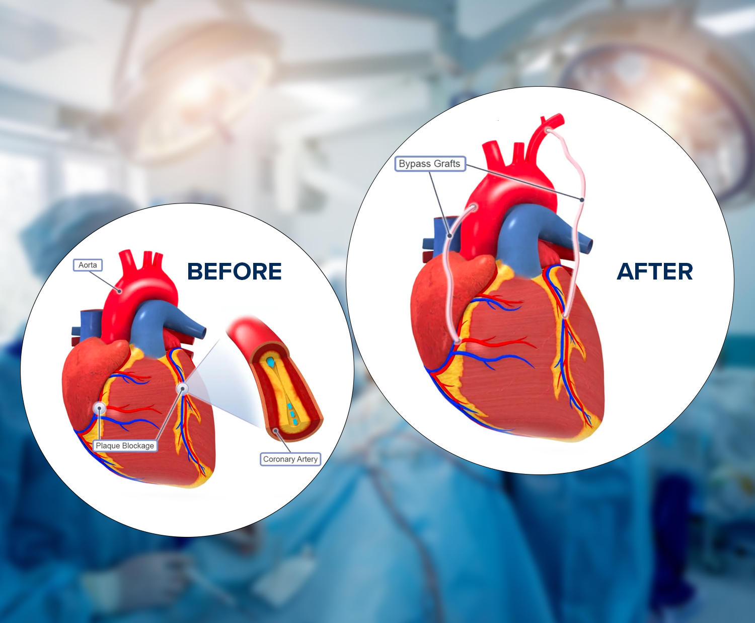
CABG Procedure Overview

Coronary Artery Bypass Grafting (CABG) is a surgical procedure commonly performed to treat coronary artery disease (CAD), a condition in which the blood vessels supplying the heart muscle (coronary arteries) become narrowed or blocked. CABG is also known as heart bypass surgery. Here is an overview of the CABG procedure:

1. Preoperative Evaluation:

Medical History and Physical Examination: A physical examination is performed after assessing the patient’s medical history and overall health.

Diagnostic Tests: These may include an electrocardiogram (ECG or EKG), an echocardiogram, coronary angiography, and other imaging tests to evaluate the extent and location of coronary artery disease.

2. Anesthesia:

Pre-anesthetic Assessment: The anesthesiologist evaluates the patient’s overall health and determines the appropriate anesthesia plan.

General Anesthesia: During surgery, the patient is put under general anesthesia, which makes them unconscious and pain-free.

3. Harvesting of Grafts:

Graft Selection: Healthy blood vessels, often taken from the patient’s own body (autografts) or occasionally from a donor (allografts), are chosen for bypass grafts. Commonly used vessels include the internal mammary artery, radial artery, and saphenous vein.

4. Surgical Access:

Sternotomy or Thoracotomy: A sternotomy (cutting through the breastbone) is the most common approach, but in some cases, a thoracotomy (a small incision between the ribs) may be used.

5. Cardiopulmonary Bypass (if needed):

Connecting to a Heart-Lung Machine: The heart may be temporarily stopped, and blood is rerouted through a heart-lung machine, providing oxygen and circulation for the body while allowing the surgeon to work on a still heart.

6. Graft Placement:

Bypassing Blockages: The surgeon attaches one end of the graft to the aorta and the other end to a coronary artery beyond the blockage, creating a new pathway for blood flow.

7. Weaning off Bypass and Closing:

Restoration of Blood Flow: If a heart-lung machine is used, the patient is gradually weaned off it, and the heart resumes pumping.

Restoration of Blood Flow: If a heart-lung machine is used, the patient is gradually weaned off it, and the heart resumes pumping.

Closure: The chest has been closed, and the incision has been sutured.

8. Postoperative Care:

Monitoring: Initially, the patient is monitored closely in the intensive care unit (ICU) before being moved to a regular room.

Recovery: Rehabilitation and recovery involve medications, lifestyle changes, and follow-up care to ensure a smooth recovery.

9. Potential Risks and Complications:

Infection, bleeding, or blood clots: As with any surgery, there are potential risks, and complications can arise. The medical team closely monitors the patient to address any issues promptly.

According to Dr. Ramji MehrotaBest Heart Specialist Doctor in India, It’s important to note that the specifics of the procedure can vary based on individual patient characteristics and the surgeon’s preference. CABG is considered a major surgery, and the decision to undergo the procedure is made after a thorough assessment of the patient’s condition and the benefits of surgery. For personalized information, it’s best to consult healthcare professionals.

It’s important to note that the specifics of the procedure can vary based on individual patient characteristics and the surgeon’s preference. CABG is considered a major surgery, and the decision to undergo the procedure is made after a thorough assessment of the patient’s condition and the benefits of surgery. For personalized information, it’s best to consult healthcare professionals.

Indications for CABG

Coronary Artery Bypass Grafting (CABG) is a surgical procedure used to treat coronary artery disease (CAD), a condition where the blood vessels that supply the heart muscle with oxygen and nutrients become narrowed or blocked. CABG is typically considered in the following indications:

  • Severe Coronary Artery Disease (CAD): CABG is often recommended when multiple coronary arteries are significantly narrowed or blocked, restricting blood flow to the heart muscle. It is a preferred option for patients with complex or diffuse CAD.
  • Angina (Chest Pain) Not Responding to Medications: When angina (chest pain) is not effectively controlled with medications or other interventions and the quality of life is significantly affected, the aim of CABG is to enhance blood flow to the heart.
  • Left Main Coronary Artery Disease: CABG is frequently recommended for patients with significant blockages in the left main coronary artery, as this vessel supplies a large portion of the heart. These blockages can pose a high risk of major cardiac events if left untreated.
  • Failed Percutaneous Coronary Intervention (PCI): If a patient has undergone PCI (angioplasty and stent placement) but experiences recurrent blockages or complications, CABG may be considered a more durable and effective solution.
  • Multi-vessel Coronary Artery Disease: CABG is often preferred for patients with blockages in multiple coronary arteries, especially if they are not amenable to PCI or if the likelihood of complete revascularization is low with angioplasty.
  • Diabetes with Coronary Artery Disease: Patients with diabetes often have more diffuse and extensive coronary artery disease. CABG may be favored over PCI in diabetic patients as it has been associated with better long-term outcomes in this population.
  • Reduced Left Ventricular Function: CABG may be considered in patients with reduced left ventricular function (ejection fraction) when medical therapy alone is insufficient to improve symptoms and prognosis.
  • Unstable Angina or Acute Coronary Syndrome: In certain cases of unstable angina or acute coronary syndrome, CABG may be performed as an emergency procedure to address severe blockages and stabilize the patient’s condition.
  • Complex Coronary Anatomy: CABG is often considered when the coronary anatomy is complex, involving multiple lesions, calcified vessels, or other factors that make PCI technically challenging or less likely to provide complete and durable revascularization.

The decision for CABG is typically made in collaboration between a cardiac surgeon, an interventional cardiologist, and other members of the heart team. It is crucial to consider the patient’s overall health, preferences, and the specific characteristics of their coronary artery disease when determining the most appropriate course of action.

What is Cardiac Rehabilitation

Cardiac rehabilitation (CR) is a structured and medically supervised program designed to help individuals recover from various heart-related conditions and improve their cardiovascular health. The primary focus of cardiac rehabilitation is to enhance the overall well-being of individuals who have experienced a heart attack, undergone heart surgery, or are living with conditions such as heart failure, coronary artery disease, or angina.

The core components of cardiac rehabilitation typically include:

  • Medical Assessment: Before starting a cardiac rehabilitation program, individuals undergo a thorough medical assessment to evaluate their current health status, identify risk factors, and create a personalized plan for rehabilitation.
  • Exercise Training: Regular, supervised exercise sessions are a fundamental aspect of cardiac rehabilitation. These sessions aim to improve cardiovascular fitness, endurance, and strength. Exercise programs are tailored to the individual’s health status, ensuring safety and gradual progression.
  • Education: Patients receive education on heart health, risk factors, lifestyle modifications, and self-management strategies. This includes information on nutrition, medication management, and stress reduction to empower individuals to make informed decisions about their health.
  • Behavioral Counseling: Psychosocial support is provided to address the emotional and psychological aspects of living with heart disease. Counseling may cover stress management, coping strategies, and mental health support.
  • Nutritional Counseling: Dietary guidance is offered to help individuals adopt heart-healthy eating habits. This may involve managing weight, reducing sodium intake, and making nutritious food choices to control factors like high blood pressure and cholesterol.
  • Risk Factor Modification: Cardiac rehabilitation emphasizes lifestyle changes to address and modify risk factors associated with heart disease. This includes smoking cessation, weight management, and strategies to control conditions like diabetes.
  • Medication Management: Education and support are provided to help individuals understand and manage their medications effectively. This includes information on medication adherence and potential side effects.
  • Monitoring and Follow-up: Regular monitoring of progress and follow-up assessments are conducted to track improvements and adjust the rehabilitation plan as needed. Follow-up care is essential to maintain and reinforce positive lifestyle changes.

According to Dr. Ramji Mehrotra, Cardiac rehabilitation is typically conducted in a specialized outpatient setting, and programs may vary in duration (typically lasting several weeks to a few months). The involvement of a multidisciplinary team, including physicians, nurses, exercise physiologists, dietitians, and psychologists, ensures a comprehensive approach to care.

Participation in cardiac rehabilitation has been associated with numerous benefits, including improved exercise capacity, reduced symptoms, lower rates of hospitalization, and enhanced quality of life for individuals with cardiovascular diseases. It plays a crucial role in the continuum of care for those recovering from heart events or managing chronic heart conditions.

Genetics and Heart Disease

Genetics and heart disease are intricately connected, and the field of study that explores this relationship is known as cardiovascular genetics. Genetics refers to the study of genes and their role in heredity and variation in living organisms. Heart disease encompasses a range of conditions that affect the heart and blood vessels, including coronary artery disease, heart failure, arrhythmias, and congenital heart defects.

Here are key points regarding genetics and heart disease:

  1. Genetic Influence: Genetic factors can contribute to an individual’s susceptibility to heart disease. Certain genetic variations may increase the risk of developing specific cardiovascular conditions.
  2. Family History: A family history of heart disease is a significant risk factor. If close relatives, such as parents or siblings, have had heart disease, heart attacks, or related conditions, it may indicate a genetic predisposition.
  3. Heritability: Heritability refers to the proportion of the variation in a trait or condition that can be attributed to genetic factors. Although genetics are important in developing heart disease, environmental and lifestyle factors also play a role.
  4. Monogenic Disorders: Mutations in a single gene can cause certain forms of heart disease. These are known as monogenic disorders. An example is familial hypercholesterolemia, a genetic condition leading to high cholesterol levels and an increased risk of atherosclerosis.
  5. Polygenic Risk: Most cases of heart disease are polygenic, meaning they result from the combined effects of multiple genetic variations. These variations may interact with each other and with environmental factors.
  6. Genetic Testing: Genetic testing can identify specific genetic variations associated with heart disease. This can be particularly useful in identifying certain hereditary conditions and guiding personalized medical management.
  7. Common Genetic Variations: Genome-wide association studies (GWAS) have identified common genetic variations associated with an increased risk of heart disease. These variations are often involved in lipid metabolism, inflammation, blood pressure regulation, and other cardiovascular processes.
  8. Gene-Environment Interaction: Genetics and environmental factors interact in influencing heart health. For example, a genetic predisposition to high cholesterol may interact with a diet high in saturated fats, leading to an increased risk of atherosclerosis.
  9. Prevention and Management: Understanding the genetic components of heart disease can contribute to more targeted prevention and management strategies. Individuals with a higher genetic risk may benefit from more vigilant monitoring and early interventions.

According to Dr. Ramji MehrotraVice Chairman – Cardiothoracic & Vascular Surgery, it’s important to note that while genetics plays a role, lifestyle factors such as diet, exercise, smoking, and stress management are crucial in preventing and managing heart disease. A comprehensive approach that considers both genetic and environmental factors is essential for understanding and addressing an individual’s risk of heart disease. Regular medical check-ups and discussions with healthcare providers can help assess and manage cardiovascular risk effectively.

Anxiety and Heart Disease

Anxiety is often considered as an emotional response to stress and has a profound impact on our physical well-being. While it is widely known that anxiety can take a toll on mental health, its intricate connection with heart disease is often underestimated. In this blog, we analyze the link between anxiety and heart disease, exploring how these seemingly unrelated facets of health are closely interlinked.

Basics of Anxiety

Anxiety is a normal response to stress, which is a built-in mechanism that helps human beings to navigate challenging situations. However, when anxiety becomes chronic or overwhelming, it can turn into a debilitating condition. Generalized Anxiety Disorder (GAD), Panic Disorder, and Social Anxiety Disorder are few of the anxiety-related conditions that affect millions of people daily.

The symptoms of anxiety can range from excessive worry and restlessness to physical manifestations like increased heart rate, muscle tension, and shortness of breath.

Connection between Anxiety and Heart Disease

Anxiety and heart disease are connected both biologically and psychologically. When we experience anxiety, our body goes into “fight or flight” mode. This response triggers the release of stress hormones like cortisol and adrenaline. While this response is essential in moments of acute danger, prolonged activation can negatively affect our cardiovascular system.

One of the immediate effects of stress hormones is an increase in heart rate and blood pressure. Over time, this can lead to a condition known as hypertension, or high blood pressure. Hypertension is a significant risk factor for heart disease. It places added strain on the heart, making it work harder and increasing the risk of various heart conditions, including coronary artery disease and heart attacks.

Moreover, chronic anxiety can lead to unhealthy lifestyle choices that further increase the risk of heart disease. Individuals with anxiety may be more likely to engage in smoking, excessive alcohol consumption, or overeating as a means of coping with their symptoms. These behaviors are all associated with an increased risk of heart disease.

Anxiety and heart disease also share a psychological connection. Living with anxiety can be very stressful, and chronic stress is detrimental to heart health. Prolonged stress can lead to unhealthy coping mechanisms, including emotional eating, lack of physical activity, obesity and social isolation.

Additionally, severe anxiety can also lead to depression. Depressive symptoms can contribute to a sedentary lifestyle and poor dietary choices, both of which are risk factors for heart disease. Moreover, individuals with anxiety and depression may have a reduced motivation to adhere to necessary heart-healthy behaviors, such as taking prescribed medications or attending medical appointments.

Prevention and Management

Some strategies that can be adopted for preventing and managing this dual threat to your health include:

  1. Stress Management: Learning effective stress management techniques, such as mindfulness, meditation, or deep breathing exercises, can help reduce the impact of anxiety on your cardiovascular system.
  2. Regular Physical Activity: Engaging in regular exercise is not only beneficial for your heart but can also help alleviate anxiety symptoms. Exercise releases endorphins, which are natural mood lifters.
  3. Healthy Diet: A balanced diet rich in fruits, vegetables, whole grains, and lean proteins can improve both your mental and heart health. Avoid excessive caffeine and sugar, as they can increase the levels of anxiety.
  4. Seek Professional Help: If anxiety is severely impacting your life, consider seeking professional help. Therapy, medication, or a combination of both can be effective in managing anxiety disorders.
  5. Medication Management: If you have existing heart conditions and are experiencing anxiety, discuss your symptoms with your doctors. They can prescribe a personalized treatment approach that considers both your heart health and anxiety.

Conclusion

The link between anxiety and heart disease highlights the importance of a holistic approach to health. Treating anxiety isn’t just about improving mental well-being; it is also about safeguarding your heart. By recognizing this connection and taking proactive steps to manage anxiety, you can reduce your risk of heart disease and live a healthier, happier life.

The link between anxiety and heart disease highlights the importance of a holistic approach to health. Treating anxiety isn’t just about improving mental well-being; it is also about safeguarding your heart. By recognizing this connection and taking proactive steps to manage anxiety, you can reduce your risk of heart disease and live a healthier, happier life.

Ventricular Tachycardia

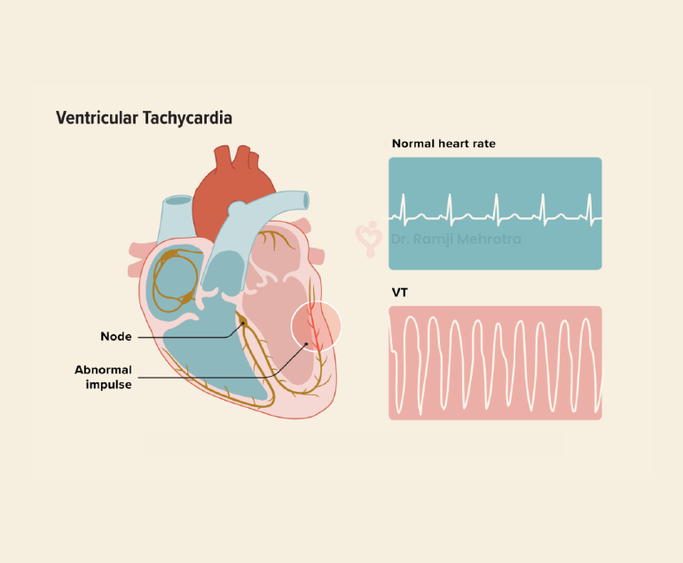
Ventricular tachycardia (VT) is a potentially life-threatening cardiac arrhythmia characterized by a rapid heartbeat originating in the ventricles, the lower chambers of the heart. In VT, the heart beats faster than the normal rate, usually over 100 beats per minute, and it can be sustained for longer periods, making it a serious medical condition that requires prompt attention and treatment.

Causes of Ventricular Tachycardia

VT typically results from abnormal electrical signals in the heart, often due to an underlying heart condition. Common causes include:

  1. Coronary Artery Disease (CAD): The most common cause of VT is CAD, which leads to insufficient blood flow to the heart muscle. This lack of oxygen can disrupt the heart’s electrical system, leading to arrhythmias like VT.
  2. Cardiomyopathy: Conditions like dilated or hypertrophic cardiomyopathy can cause changes in the heart’s structure and electrical system, increasing the risk of VT.
  3. Heart Attack: An acute myocardial infarction (heart attack) can scar heart tissue, creating areas where abnormal electrical impulses can arise.
  4. Valvular Heart Disease: Malfunctioning heart valves can affect blood flow, increasing the likelihood of arrhythmias, including VT.
  5. Electrolyte Imbalances: Abnormal levels of electrolytes (such as potassium, sodium, or calcium) can disrupt the heart’s electrical signals.

Symptoms

The symptoms of VT can vary in severity and may include:

  • Palpitations: A strong, rapid heartbeat is a hallmark symptom of VT.
  • Chest Pain: Some people may experience chest discomfort or pain during VT episodes.
  • Dizziness or Lightheadedness: Insufficient blood flow to the brain can lead to these sensations.
  • Shortness of Breath: Rapid heartbeats can reduce the heart’s efficiency, leading to breathlessness.
  • Loss of Consciousness: In severe cases, VT can cause fainting or even sudden cardiac arrest.

Diagnosis

A proper diagnosis of VT involves a combination of medical history, physical examination, and diagnostic tests, including:

  1. Electrocardiogram (ECG or EKG): This test records the heart’s electrical activity and can identify VT patterns.
  2. Holter Monitor: A portable ECG device worn for an extended period can capture intermittent VT episodes.
  3. Electrophysiology Study (EPS): Invasive testing, where thin wires are inserted into the heart, can pinpoint the location and cause of VT.
  4. Echocardiogram: This ultrasound imaging of the heart can reveal structural abnormalities.

Treatment Options

The management of VT depends on its cause, severity, and the patient’s overall health. Treatment options may include:

  1. Medications: Antiarrhythmic drugs can help control VT episodes by stabilizing the heart’s electrical activity.
  2. Implantable Cardioverter-Defibrillator (ICD): For those at high risk of life-threatening VT, an ICD is surgically implanted to monitor and correct abnormal rhythms by delivering shocks when needed.
  3. Catheter Ablation: This procedure involves the use of catheters to target and destroy the abnormal heart tissue responsible for VT.
  4. Lifestyle Changes: Managing underlying conditions like CAD or cardiomyopathy through lifestyle modifications, such as diet, exercise, and smoking cessation, can reduce the risk of VT.
  5. Emergency Care: If VT leads to cardiac arrest, immediate defibrillation is crucial to restore normal rhythm and save a life.

Conclusion

Ventricular tachycardia is a serious cardiac arrhythmia that requires careful evaluation and management. Early diagnosis and appropriate treatment are essential to reduce the risk of life-threatening complications. Patients with a history of heart disease or risk factors should consult with the doctors regularly for them to monitor and address any cardiac issues, ultimately improving their overall heart health and quality of life.

Cardiovascular Conditions in Older Patients

Cardiovascular conditions in older patients pose significant challenges due to the natural aging process and the increased prevalence of risk factors associated with advanced age. As individuals grow older, their cardiovascular system undergoes changes that can increase the likelihood of developing various heart-related disorders.

Some of the common cardiovascular conditions seen in older patients include:

Coronary Artery Disease (CAD)

One of the most prevalent cardiovascular conditions in older adults is coronary artery disease (CAD). CAD occurs when the arteries that supply blood to the heart become narrowed or blocked due to atherosclerosis, the build-up of plaque. Older adults are more at risk to CAD due to factors such as prolonged exposure to risk factors like high blood pressure, high cholesterol, and smoking.

Diagnosis of CAD in older patients may be challenging because symptoms can be masked by other age-related conditions. Doctors employ diagnostic tools such as stress tests and cardiac imaging to confirm the diagnosis. Treatment typically involves a combination of lifestyle modifications, medication management, and, in severe cases, invasive interventions like angioplasty or bypass surgery.

Heart failure

Heart failure is another common cardiovascular condition in older patients. It occurs when the heart is unable to pump blood efficiently, leading to symptoms such as shortness of breath, fatigue, and fluid retention. Age-related changes in the heart, such as reduced elasticity and stiffening of the cardiac muscles, increase the risk of heart failure.

Accurate diagnosis of heart failure requires a comprehensive assessment of symptoms, physical examination, and diagnostic tests like echocardiography. Treatment involves a combination of medications to manage symptoms and improve heart function, lifestyle modifications like fluid and salt restriction, and, in some cases, devices like pacemakers or implantable defibrillators.

Arrhythmias

Arrhythmias, including atrial fibrillation (AF), are commonly encountered in older adults. AF, characterized by irregular and rapid heartbeat, is associated with an increased risk of stroke, heart failure, and other cardiovascular complications.

Advanced age is a significant risk factor for developing AF due to changes in the electrical conduction system of the heart. Diagnosis involves an electrocardiogram (ECG) and may require additional monitoring methods like Holter monitoring or event recorders. Management of AF in older patients aims to control heart rate, prevent blood clots with anticoagulant therapy, and restore normal heart rhythm when possible.

Hypertension

Hypertension, or high blood pressure, is highly prevalent in older adults and is a major risk factor for cardiovascular disease. Age-related changes in blood vessels, hormonal factors, and lifestyle habits contribute to the development of hypertension.

Effective management of hypertension in older patients involves lifestyle modifications, such as a heart-healthy diet and regular exercise, along with medications to achieve blood pressure targets. Close monitoring and regular follow-up are essential to ensure blood pressure control and prevent complications.

Conclusion

Cardiovascular conditions in older patients require a comprehensive and individualized approach to diagnosis and treatment. Age-related changes in the cardiovascular system increase the vulnerability of older adults to conditions like coronary artery disease, heart failure, arrhythmias, and hypertension. Addressing modifiable risk factors and promoting healthy lifestyle habits are essential for preventing and managing cardiovascular conditions in older patients.

Ross Procedure

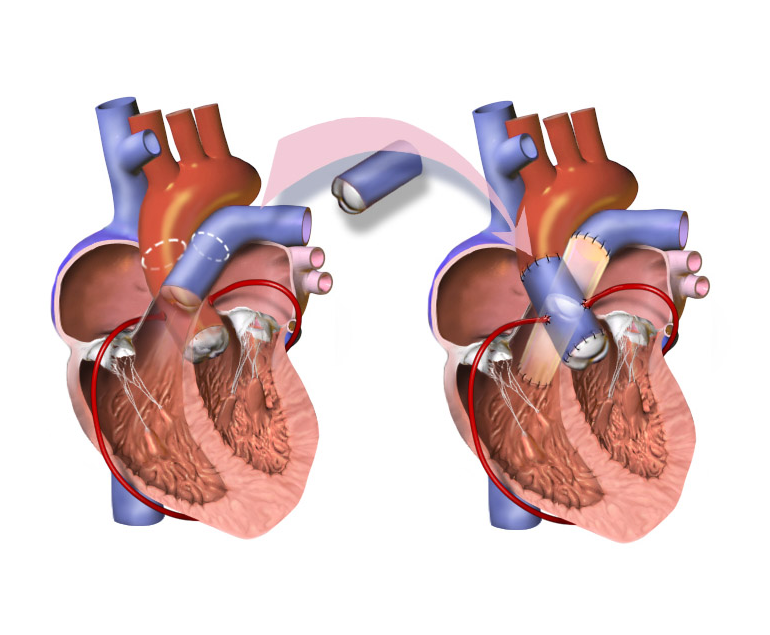
The Ross procedure is a surgical procedure used to treat aortic valve disease, which is a condition in which the valve that controls blood flow from the heart to the aorta becomes narrowed or leaks. This procedure involves replacing the damaged aortic valve with the patient’s own pulmonary valve, and then replacing the pulmonary valve with a donated or artificial valve.

The Ross procedure is named after its creator, Donald Ross, a Scottish cardiac surgeon who developed the technique in the 1960s. Since then, the procedure has become a popular option for treating aortic valve disease in certain patients.

Advantages of Ross Procedure

The Ross procedure is typically recommended for younger patients who have aortic valve disease, as it has several advantages over other types of valve replacement surgery. One of the main advantages is that it avoids the need for lifelong anticoagulation therapy, which is required for patients who receive a mechanical valve replacement. Anticoagulation therapy is a type of medication that helps prevent blood clots, but it can also increase the risk of bleeding and other complications.

Another advantage of the Ross procedure is that it provides a better quality of life for patients. The pulmonary valve is better suited to the low-pressure environment of the aortic position than artificial valves, which can often lead to complications such as valve dysfunction or blood clots.

Procedure

The Ross procedure is performed under general anaesthesia and typically takes many hours to complete. During the procedure, the surgeon will make an incision in the chest and access the heart. The patient’s pulmonary valve will then be removed and used to replace the damaged aortic valve. A new pulmonary valve, either from a donated cadaver or an artificial valve, will then be used to replace the removed pulmonary valve.

After the procedure, patients may need to spend several days in the hospital recovering. They may experience some discomfort or pain, and will need to take medication to manage pain and prevent infection. Patients will also need to gradually increase their physical activity and follow a prescribed rehabilitation program to help them recover.

Suitability

While the Ross procedure has many benefits, it is not suitable for all patients with aortic valve disease. Patients who have other heart conditions, such as coronary artery disease or an enlarged heart, may not be good candidates for the procedure. Additionally, patients who have already had a previous valve replacement or who have other medical conditions that could complicate surgery may not be good candidates for the procedure.

Conclusion

Ross procedure is a surgical procedure used to treat aortic valve disease in certain patients. It involves replacing the damaged aortic valve with the patient’s own pulmonary valve, and then replacing the pulmonary valve with a donated or artificial valve. If you are considering the Ross procedure, it is important to discuss the risks and benefits with your doctor to determine if it is the right option for you.

What is Coronary Artery Disease?

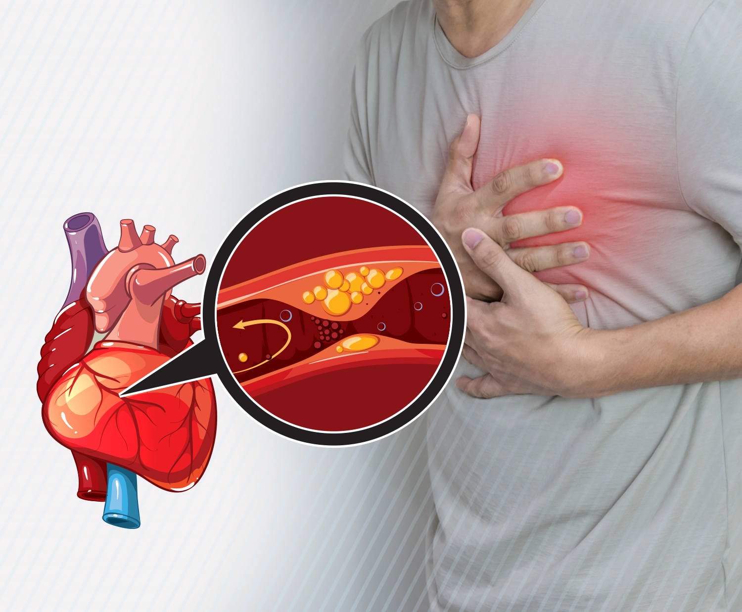
Coronary artery disease (CAD) or coronary heart disease is a common type of heart disease that occurs when the arteries become hardened and narrowed. This is caused due to the cholesterol and calcium deposit or plaque in the arteries.

Coronary artery disease begins due to a condition called atherosclerosis which occurs when cholesterol and calcium collects on the inner walls of the arteries. This build-up is known as plaque which can block blood flow due to the narrowing of the arteries. If the plaque bursts it can lead to a blood clot.

Symptoms of CAD

A patient having coronary artery disease can have chest pain, breathlessness, fatigue, nausea and due to the reduced blood flow to the heart. Chest discomfort or angina is accompanied by symptoms like numbness, heaviness, aching, squeezing, etc.

Complete blockage of blood flow can also lead to heart attack.

Risk Factors

In addition to high cholesterol, other causes of damage to coronary arteries include high blood pressure, diabetes, smoking, sedentary lifestyle, etc.

Obesity, stress, unhealthy diet, irregular sleep and alcohol usage are other risk factors for coronary artery disease.

Diagnosis of CAD

The doctor reviews the patient’s symptoms, risk factors and medical history before performing a physical examination. Diagnostic tests include:

  • Electrocardiograph test: This test can detect heart attack and heart rhythm issues by recording the heart’s electrical activity.
  • Exercise stress test: This is a treadmill test that can help suspect  coronary blockages
  • Pharmacologic stress test: This test can also help detect coronary blockages through medication given to increase heart rate. This is done in patients who cannot walk on treadmill.
  • Coronary calcium scan: This can identify the amount of calcium on the walls of the coronary arteries which can give an indication of atherosclerosis.
  • Echocardiogram: This test utilizes sound waves to measure the overall functioning of the heart.
  • Blood tests: These tests are done for factors affecting arteries like cholesterol, triglycerides, lipoprotein, glucose, etc.
  • Cardiac catheterization: In this test small tubes are inserted into the blood vessels to detect the presence of coronary artery disease and also evaluate the heart function.

Prevention of CAD

According to Dr. Ramji Mehrotra who is one of India’s leading cardiologist, coronary artery disease can be prevented by adopting a healthy lifestyle to improve heart health. These include stopping smoking and limiting alcohol use, consuming a heart-healthy diet, exercising and increasing activity levels, etc.

Blood pressure, diabetes and cholesterol should be controlled. Stress management is also essential to prevent coronary artery disease.