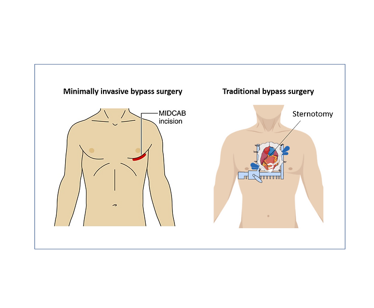
Silent heart attack: What are the risks?

A silent heart attack, medically termed a “silent myocardial infarction,” is a potentially dangerous event that often goes unnoticed due to its lack of typical symptoms. Unlike a traditional heart attack, which is accompanied by chest pain, shortness of breath, and discomfort in the arms, neck, jaw, or back, a silent heart attack occurs without these apparent signs. This makes it a significant concern, as individuals are less likely to seek immediate medical attention, leading to delayed diagnosis and treatment. Understanding the risks associated with silent heart attacks is crucial for prevention and proactive healthcare management.

Causes and Risks

Silent heart attacks share the same underlying cause as symptomatic heart attacks: a disruption of blood flow to a portion of the heart muscle due to a blocked or narrowed coronary artery. This interruption leads to the death of heart muscle cells, which can impair cardiac function and increase the risk of future heart-related complications. The factors that contribute to silent heart attacks are often the same as those for traditional heart attacks, including high blood pressure, high cholesterol, diabetes, obesity, smoking, and a sedentary lifestyle. However, certain demographic groups, such as older adults and individuals with diabetes, are more prone to experiencing silent heart attacks.

One of the major dangers of silent heart attacks is their gradual progression. Without the characteristic chest pain that typically prompts individuals to seek medical help, silent heart attacks can go undetected for weeks, months, or even years. During this time, untreated heart damage may accumulate, putting individuals at greater risk of heart failure, abnormal heart rhythms (arrhythmias), and subsequent heart attacks. Additionally, silent heart attacks are often identified incidentally when a person undergoes medical tests for unrelated issues, further highlighting the hidden nature of these events.

Preventive Measures

The lack of awareness surrounding silent heart attacks highlights the importance of preventive healthcare measures. Regular check-ups and screenings are essential, especially for individuals with risk factors. Blood pressure, cholesterol, and blood sugar levels should be closely monitored, and lifestyle modifications should be adopted to mitigate these risk factors. Adopting a heart-healthy diet rich in fruits, vegetables, whole grains, lean proteins, and healthy fats, along with engaging in regular physical activity, can significantly reduce the likelihood of both silent and symptomatic heart attacks.

Moreover, recognizing potential warning signs that might not be as dramatic as traditional symptoms is vital. Unexplained fatigue, mild discomfort in the chest, nausea, or discomfort in the upper abdomen, back, or jaw could all be indicative of a silent heart attack. Hence, they should not be ignored, especially if they persist or worsen over time.

Conclusion

Silent heart attacks pose a grave risk due to their inconspicuous nature and delayed diagnosis. The absence of classic symptoms can lead to untreated heart damage and increase the likelihood of complications. It is essential for individuals, particularly those with risk factors, to prioritize regular health check-ups, maintain a heart-healthy lifestyle, and be vigilant about any unusual or persistent symptoms. By taking proactive measures, individuals can mitigate the risks associated with silent heart attacks and promote overall heart health.

Cardiac Amyloidosis

Cardiac amyloidosis is a rare and potentially fatal disorder that arises from the abnormal accumulation of amyloid protein in the heart tissues, leading to compromised cardiac function.

Nature of Cardiac Amyloidosis

Cardiac amyloidosis stems from the buildup of amyloid fibrils, which are insoluble protein aggregates that accumulate in various organs, including the heart. These amyloid deposits infiltrate the heart muscles, causing them to become stiff, thickened, and less compliant. Over time, this impairs the heart’s ability to pump blood effectively, leading to heart failure and other cardiovascular complications.

Subtypes of Cardiac Amyloidosis

There are different subtypes of cardiac amyloidosis, each originating from distinct precursor proteins. The two most prevalent subtypes are:

  • AL Amyloidosis (Immunoglobulin Light Chain Amyloidosis): This form arises when abnormal plasma cells in the bone marrow produce excessive amounts of light chains, a component of antibodies. These light chains then accumulate as amyloid fibrils in various organs, including the heart.
  • ATTR Amyloidosis (Transthyretin Amyloidosis): In ATTR amyloidosis, the precursor protein is transthyretin, a protein responsible for transporting thyroid hormone and retinol. Mutations or aging-related changes in transthyretin can lead to the formation of amyloid fibrils that deposit in the heart.

Challenges in Diagnosis

Cardiac amyloidosis remains a diagnostic challenge due to its overlapping symptoms with other heart conditions and its rarity. Common symptoms include fatigue, shortness of breath, swelling in the legs, and irregular heartbeats. These nonspecific signs often delay accurate diagnosis, which is further complicated by the fact that cardiac amyloidosis can coexist with other heart diseases.

Advancements in Detection

Recent advancements in imaging techniques have greatly improved the detection of cardiac amyloidosis. Cardiac MRI, nuclear imaging with bone tracers, and echocardiography can provide insights into the presence and extent of amyloid deposits in the heart. Additionally, advances in genetic testing help in identifying ATTR amyloidosis, enabling early intervention for individuals with genetic predisposition.

Management and Treatment

Managing cardiac amyloidosis involves addressing both the underlying cause and the associated symptoms. In AL amyloidosis, treatments focus on suppressing the abnormal plasma cells responsible for producing the amyloidogenic light chains. Chemotherapy and stem cell transplantation are commonly used in the treatment. For ATTR amyloidosis, treatment options include stabilizing the transthyretin protein or, in cases of hereditary ATTR, gene-silencing therapies.

Heart Disease and Pericardiocentesis

Heart disease is one of the leading causes of mortality and requires advanced medical interventions. Pericardiocentesis is a critical procedure in addressing complications related to pericardial effusion – a condition characterized by the accumulation of fluid in the pericardium, the protective sac around the heart. This procedure plays a vital role in both diagnosing and treating underlying heart conditions, alleviating symptoms, and preventing potentially life-threatening complications.

Causes

Pericardial effusion can result from various factors such as infections, inflammation, malignancies, trauma, or as a consequence of cardiac surgery. The accumulation of excess fluid within the pericardial space can exert pressure on the heart, impairing its ability to pump blood effectively. This can lead to symptoms like chest pain, shortness of breath, and even cardiac tamponade – a condition where the pressure from the fluid buildup compresses the heart chambers, compromising its function and circulation.

Procedure

Pericardiocentesis involves the insertion of a needle or catheter into the pericardial space to drain the accumulated fluid. This procedure serves a dual purpose: providing symptomatic relief by relieving the pressure on the heart and facilitating diagnostic evaluation by analyzing the removed fluid for potential underlying causes.

The pericardiocentesis procedure is performed under imaging guidance, such as echocardiography or fluoroscopy, to ensure precision and minimize the risk of complications. The needle is carefully guided into the pericardial sac, and the fluid is aspirated. In some cases, a catheter may be left in place for continuous drainage if there is a significant fluid buildup or if the effusion is recurrent.

Benefits

Pericardiocentesis can provide rapid relief from the debilitating symptoms caused by pericardial effusion. By promptly reducing the pressure on the heart, patients often experience a marked improvement in their ability to breathe and reduced discomfort. Additionally, the removed fluid can be sent for analysis, which helps in diagnosing the underlying cause of the effusion. This diagnostic insight is crucial in guiding subsequent treatment decisions.

In some cases, pericardiocentesis may serve as a temporary measure while other interventions, such as medication, are initiated to address the underlying condition causing the effusion. However, in more severe cases where pericardial effusion poses an immediate threat to cardiac function, pericardiocentesis can be a life-saving intervention.

Conclusion

Pericardiocentesis plays a vital role in the management of heart disease, specifically in addressing the complications arising from pericardial effusion. This procedure offers both diagnostic and therapeutic benefits, helping in the swift mitigation of symptoms and potentially preventing life-threatening cardiac complications.

Dilated Cardiomyopathy

Dilated cardiomyopathy (DCM) is a condition characterized by the dilation and weakening of the heart’s main pumping chamber, the left ventricle. This chronic and progressive disorder affects the heart muscle, leading to impaired cardiac function and potentially causing heart failure. DCM can also affect the right ventricle and both ventricles in some cases.

Causes

The exact causes of DCM are often unknown, but various factors can contribute to its development. These include genetic mutations, viral infections (such as viral myocarditis), exposure to toxins (for example alcohol), autoimmune diseases, and metabolic disorders. In many cases, DCM is considered idiopathic, meaning the cause is uncertain.

Symptoms

DCM can present with a range of symptoms, which may include fatigue, shortness of breath, swelling of the legs and ankles (edema), rapid or irregular heartbeat (arrhythmias), chest pain, dizziness, and fainting. These symptoms are often related to the heart’s inability to effectively pump blood and supply oxygen to the body’s tissues.

Diagnosis

To diagnose DCM, a comprehensive evaluation is required, which typically involves a thorough medical history review, physical examination, and various diagnostic tests. These tests may include electrocardiogram (ECG), echocardiogram (to assess heart structure and function), cardiac MRI or CT scan (for detailed imaging), blood tests (to rule out other conditions), and occasionally a heart biopsy.

Treatment

The management of DCM aims to improve symptoms, slow disease progression, and prevent complications. Treatment approaches may include:

  1. Medications: Several medications can be prescribed to manage symptoms, reduce the workload on the heart, control blood pressure, prevent blood clots, and treat arrhythmias. These may include beta-blockers, ACE inhibitors, diuretics, anticoagulants, and anti-arrhythmic drugs.
  • Lifestyle modifications: Patients need to make lifestyle changes like reducing salt intake, avoiding alcohol, stopping smoking, maintaining proper weight, and exercising as required.
  • Device therapy: In advanced cases or those with severe heart failure, implantable devices like pacemakers or defibrillators may be recommended to regulate the heart’s rhythm or deliver electrical shocks if life-threatening arrhythmias occur.
  • Cardiac rehabilitation: This supervised program combines exercise, education, and counseling to help patients recover and improve their overall cardiovascular health.
  • Surgical interventions: In some cases, surgical interventions may be necessary, such as coronary artery bypass grafting (CABG) to improve blood flow to the heart, or heart transplantation for end-stage heart failure.

Prognosis

The prognosis for individuals with DCM can vary widely depending on the underlying cause, severity of symptoms, and response to treatment. With appropriate management, lifestyle modifications, and adherence to medication, many patients can experience an improved quality of life and a slower disease progression. However, some individuals may develop more severe heart failure and require advanced therapies or heart transplantation. Regular follow-up with a cardiologist is essential to monitor the condition and adjust treatment as needed.

Conclusion

Dilated cardiomyopathy is a complex heart condition characterized by the enlargement and weakening of the heart muscle. While the exact causes are unclear in many cases, a combination of genetic, viral, autoimmune, and environmental factors can contribute to its development. Early diagnosis, appropriate medical management, lifestyle modifications, and sometimes surgical interventions are essential for managing the symptoms, slowing disease progression, and improving the prognosis for individuals with DCM.

Ross Procedure

The Ross procedure is a surgical procedure used to treat aortic valve disease, which is a condition in which the valve that controls blood flow from the heart to the aorta becomes narrowed or leaks. This procedure involves replacing the damaged aortic valve with the patient’s own pulmonary valve, and then replacing the pulmonary valve with a donated or artificial valve.

The Ross procedure is named after its creator, Donald Ross, a Scottish cardiac surgeon who developed the technique in the 1960s. Since then, the procedure has become a popular option for treating aortic valve disease in certain patients.

Advantages of Ross Procedure

The Ross procedure is typically recommended for younger patients who have aortic valve disease, as it has several advantages over other types of valve replacement surgery. One of the main advantages is that it avoids the need for lifelong anticoagulation therapy, which is required for patients who receive a mechanical valve replacement. Anticoagulation therapy is a type of medication that helps prevent blood clots, but it can also increase the risk of bleeding and other complications.

Another advantage of the Ross procedure is that it provides a better quality of life for patients. The pulmonary valve is better suited to the low-pressure environment of the aortic position than artificial valves, which can often lead to complications such as valve dysfunction or blood clots.

Procedure

The Ross procedure is performed under general anaesthesia and typically takes many hours to complete. During the procedure, the surgeon will make an incision in the chest and access the heart. The patient’s pulmonary valve will then be removed and used to replace the damaged aortic valve. A new pulmonary valve, either from a donated cadaver or an artificial valve, will then be used to replace the removed pulmonary valve.

After the procedure, patients may need to spend several days in the hospital recovering. They may experience some discomfort or pain, and will need to take medication to manage pain and prevent infection. Patients will also need to gradually increase their physical activity and follow a prescribed rehabilitation program to help them recover.

Suitability

While the Ross procedure has many benefits, it is not suitable for all patients with aortic valve disease. Patients who have other heart conditions, such as coronary artery disease or an enlarged heart, may not be good candidates for the procedure. Additionally, patients who have already had a previous valve replacement or who have other medical conditions that could complicate surgery may not be good candidates for the procedure.

Conclusion

Ross procedure is a surgical procedure used to treat aortic valve disease in certain patients. It involves replacing the damaged aortic valve with the patient’s own pulmonary valve, and then replacing the pulmonary valve with a donated or artificial valve. If you are considering the Ross procedure, it is important to discuss the risks and benefits with your doctor to determine if it is the right option for you.

What is Cardiac Catheterization?

Cardiac catheterization, also known as coronary angiography, is a medical procedure that allows doctors to examine the heart and blood vessels to identify any problems. It is a minimally invasive procedure that involves inserting a thin, flexible tube called a catheter into a blood vessel, typically in the groin or arm, and guiding it through the blood vessels to the heart.

The catheterization procedure is performed in a cardiac catheterization laboratory (also known as a cath lab) by a cardiologist. The procedure is typically done under local anaesthesia, which numbs the area where the catheter will be inserted.

Procedure

During the procedure, the cardiologist will make a small incision in the skin and insert a hollow sheath into the blood vessel. The catheter is then inserted through the sheath and guided to the heart using x-ray guidance. Once the catheter is in place, the cardiologist can inject a contrast dye into the bloodstream, which makes the heart and blood vessels visible on x-ray.

The contrast dye allows the cardiologist to examine the heart and blood vessels for any abnormalities or blockages that may be present. If a blockage is found, the cardiologist can perform a procedure called angioplasty to open the blocked artery and restore blood flow to the heart.

Diagnostic Tool

Cardiac catheterization is a valuable diagnostic tool that can provide important information about the heart and blood vessels. It is typically used to diagnose and evaluate a range of heart conditions, including coronary artery disease, heart valve problems, congenital heart defects, and heart failure.

In addition to providing diagnostic information, cardiac catheterization can also be used to treat certain heart conditions. For example, angioplasty can be used to open blocked arteries and restore blood flow to the heart. Other procedures, such as stent placement and balloon valvuloplasty, can also be performed during cardiac catheterization.

Risks

While cardiac catheterization is generally considered a safe procedure, there are some risks associated with it. These include bleeding, infection, damage to the blood vessels, and allergic reactions to the contrast dye. However, these risks are generally low and can be minimized with proper preparation and monitoring.

Tests before the procedure

Before undergoing cardiac catheterization, patients will typically undergo a series of tests to evaluate their heart health and determine if they are a good candidate for the procedure. These tests may include an electrocardiogram (ECG), echocardiogram, stress test, and blood tests.

Patients should also inform their doctor of any medications they are taking, as some medications may need to be temporarily stopped prior to the procedure. Patients should also avoid eating or drinking anything for several hours before the procedure.

Conclusion

Overall, cardiac catheterization is a valuable tool for diagnosing and treating a range of heart conditions. While there are some risks associated with the procedure, these risks are generally low and can be minimized with proper preparation and monitoring. Patients should talk to their doctor about whether cardiac catheterization is a good option for them and what they can expect during and after the procedure.

Electrical Cardioversion

Electrical cardioversion is a medical procedure that is used to treat abnormal heart rhythms, also known as arrhythmias. This procedure involves the delivery of a controlled electric shock to the heart through two large pads or paddles placed on the chest. The shock interrupts the abnormal electrical activity of the heart and allows it to reset to its normal rhythm.

Arrhythmias

Arrhythmias can occur in the upper chambers of the heart (atria) or the lower chambers (ventricles). The most common arrhythmias are atrial fibrillation, atrial flutter, and supraventricular tachycardia. These arrhythmias can cause symptoms such as palpitations, dizziness, shortness of breath, chest pain, and fatigue. If left untreated, they can also increase the risk of stroke, heart failure, and other serious complications.

Electrical cardioversion procedure

Electrical cardioversion is performed by a trained healthcare provider such as a cardiologist. Before the procedure, the patient is given medications to reduce the risk of blood clots and to ensure that the heart is in the correct rhythm. The cardiologist then places the pads or paddles on the chest of the patient, and a brief electric shock is delivered to the heart.

The amount of electricity delivered is carefully calibrated to ensure that it is enough to reset the heart’s rhythm without causing any damage to the heart or surrounding tissues. The shock can cause the patient’s muscles to contract, which can sometimes be painful. However, patients are typically given sedation or anaesthesia to minimize discomfort during the procedure.

Once the heart has been reset to its normal rhythm, the patient is monitored closely for a period of time to ensure that the heart remains stable. In some cases, additional electrical shocks may be needed to maintain the normal rhythm.

Electrical cardioversion is a highly effective procedure for treating certain types of arrhythmias. In many cases, it can restore normal heart function almost immediately. However, the success of the procedure depends on several factors, including the type and severity of the arrhythmia, the duration of the arrhythmia, and the overall health of the patient.

Medical evaluation before electrical cardioversion

Before undergoing electrical cardioversion, patients will usually undergo a thorough medical evaluation to ensure that they are good candidates for the procedure. This evaluation may include blood tests, an electrocardiogram (ECG), a chest X-ray, and other tests as needed. The healthcare provider will also review the patient’s medical history, including any medications or supplements that the patient is taking.

Patients with certain conditions may not be suited for electrical cardioversion. For example, patients with certain types of heart disease, severe lung disease, or a history of blood clots may be at increased risk of complications from the procedure. The doctor will carefully evaluate the patient’s medical history and perform any necessary tests to determine whether electrical cardioversion is safe and appropriate.

Electrical cardioversion is generally a safe procedure, but like any medical procedure, it can have some risks. The most common side effect is mild skin irritation or burns where the pads or paddles are placed on the chest. These typically heal within a few days.

Patients may also be advised to make certain lifestyle changes, such as quitting smoking, reducing alcohol intake, and losing weight, to reduce the risk of future arrhythmias.

Conclusion

Electrical cardioversion is an effective treatment for certain types of arrhythmias. With proper care and follow-up, many patients can expect to have improved heart function and reduced risk of future arrhythmias.

Complete Atrioventricular Canal Defect (CAVC)

Complete atrioventricular canal defect (CAVC) is a rare congenital heart condition that occurs when there is a hole in the centre of the heart that affects the atria and ventricles. This condition is also known as an atrioventricular septal defect (AVSD), and it can cause a range of serious health problems if left untreated.

In a normal heart, there is a wall (septum) between the two atria and another wall between the two ventricles. These walls prevent the mixing of oxygen-rich and oxygen-poor blood. In CAVC, these walls are missing, and a large hole exists between the atria and ventricles. As a result, oxygen-rich blood mixes with oxygen-poor blood, leading to reduced oxygen levels in the body and heart failure over time.

Diagnosis of CAVC

CAVC is typically diagnosed in infancy or early childhood. Infants with CAVC may experience symptoms such as difficulty breathing, rapid breathing, and feeding difficulties. In some cases, babies with CAVC may also have low oxygen levels, which can cause a bluish tint to the skin (cyanosis).

The diagnosis of CAVC is usually confirmed through diagnostic tests such as an echocardiogram, which uses ultrasound waves to create images of the heart. Other tests that may be performed include an electrocardiogram (ECG) to evaluate the heart’s electrical activity and a chest X-ray to check for signs of heart enlargement.

Treatment

Treatment for CAVC usually involves surgery to close the hole in the heart and reconstruct the affected valves. This type of surgery is typically done in early infancy or early childhood, depending on the severity of the condition. In some cases, medication may be prescribed to manage symptoms such as heart failure or to prevent complications such as infection.

Long-term outlook

The long-term outlook for children with CAVC depends on the severity of the defect and the timing of treatment. In general, children who receive timely and appropriate treatment have a good prognosis and can live normal, healthy lives. However, in severe cases, CAVC can lead to heart failure, pulmonary hypertension, and other serious complications.

In addition to medical treatment, children with CAVC may require ongoing monitoring and care throughout their lives. This may include regular check-ups with a pediatric cardiologist, medication management, and follow-up diagnostic tests such as echocardiograms.

Conclusion

Complete atrioventricular canal defect is a rare but serious congenital heart condition that affects the atria and ventricles of the heart. Early diagnosis and appropriate treatment are crucial for managing the condition and preventing complications. If you suspect your child may have CAVC, it is important to seek medical attention promptly to ensure the best possible outcome.

How Positron Emission Tomography (PET) Scan Works?

Positron emission tomography (PET) is a medical imaging technique that produces detailed images of the inside of the body. It is used to diagnose and monitor various medical conditions, including cancer, heart disease, and neurological disorders.

What is a PET scan?

A PET scan is a type of medical imaging that uses a small amount of a radioactive substance, called a tracer, to produce images of the inside of the body. The tracer is injected into the body, swallowed, or inhaled, and then detected by a PET scanner. The PET scanner is a large machine that contains a ring of detectors that can detect the radiation emitted by the tracer.

How does a PET scan work?

The tracer used in a PET scan is typically a compound that contains a small amount of a radioactive substance, such as fluorine-18 or carbon-11. These radioactive substances emit positrons, which are positively charged particles that are the opposite of electrons. When a positron encounters an electron in the body, they annihilate each other and emit two gamma rays in opposite directions.

The PET scanner detects the gamma rays and uses them to create a three-dimensional image of the area being scanned. The scanner can detect the gamma rays from multiple angles, which allows it to create a detailed image of the area.

What happens during a PET scan?

Before the PET scan, the patient is injected with the tracer. The tracer is typically injected into a vein in the arm, but it can also be swallowed or inhaled, depending on the type of scan being done.

After the tracer is injected, the patient is asked to rest for a period of time to allow the tracer to circulate through the body. The amount of time the patient needs to rest depends on the type of scan being done and the tracer being used.

Once the tracer has circulated through the body, the patient is taken to the PET scanner. The scanner is a large machine with a narrow table that slides in and out of the machine.

The patient lies on the table, and the scanner is positioned over the area being scanned. The patient needs to remain still during the scan to ensure the images are clear.

The scanner emits a small amount of radiation to detect the tracer in the body. The patient will not feel anything during the scan, but they may hear a clicking or buzzing sound coming from the machine.

After the scan is complete, the patient can go home. The amount of radiation in the tracer is small, and it typically does not pose a health risk.

What are the uses of PET scans?

PET scans are used to diagnose and monitor a variety of medical conditions, including:

  1. Cancer: PET scans can help to diagnose cancer and determine the extent of the disease. They can also be used to monitor the effectiveness of cancer treatments, such as chemotherapy.
  2. Heart disease: PET scans can be used to diagnose heart disease and determine the extent of damage to the heart muscle. They can also be used to assess blood flow to the heart and to determine the effectiveness of treatments, such as angioplasty.
  3. Neurological disorders: PET scans can be used to diagnose and monitor neurological disorders, such as Alzheimer’s disease, Parkinson’s disease, and epilepsy.
  4. Mental health disorders: PET scans can be used to study the brain and to diagnose and monitor mental health disorders, such as depression and schizophrenia.

Different Types of Heart Disease

Heart disease, also known as cardiovascular disease, refers to a range of conditions that affect the heart and blood vessels. The heart is a vital organ that pumps blood throughout the body, and its proper functioning is essential for good health. However, various factors such as genetics, lifestyle choices, and other health conditions can contribute to the development of heart disease. In this blog we will discuss different types of heart diseases and how they affect the heart and blood vessels.

  • Coronary Artery Disease (CAD)

Coronary artery disease is a condition that occurs when the blood vessels that supply blood to the heart become narrow or blocked, causing a reduction in blood flow to the heart muscle especially when demand is high on exertion. This can lead to chest pain, angina, and heart attack. The primary cause of CAD is the build-up of plaque in the coronary arteries. Plaque is a mixture of cholesterol, fat, and other substances that can clog the arteries and make it difficult for blood to flow. The risk factors for CAD include high cholesterol, high blood pressure, smoking, obesity, and a family history of heart disease.

  • Heart Attack

A heart attack, also known as myocardial infarction, occurs when a blood clot or plaque ruptures in a coronary artery, blocking blood flow to a portion of the heart muscle. This can cause damage or death to the heart muscle, which can lead to serious health problems, including heart failure. The symptoms of a heart attack include chest pain, shortness of breath, sweating, and light headedness. Quick treatment with medications and procedures such as angioplasty or bypass surgery can help prevent further damage to the heart and reduce the risk of heart failure.

  • Heart Failure

During heart failure the heart is unable to pump sufficient blood to meet the body’s requirements. This can occur due to damage to the heart muscle from a heart attack, heart valve dysfunction, myopathy, certain infections, high blood pressure, or other health conditions. Heart failure can cause a number of symptoms, including fatigue, shortness of breath, and swelling in the legs, ankles, and feet. Treatment for heart failure includes medications, lifestyle changes, and in severe cases, surgery to implant a pacemaker or defibrillator, ventricular assist devices and heart transplant.

  • Arrhythmia

Arrhythmia is an irregular heartbeat that can occur due to a variety of factors, including heart disease, high blood pressure, and stress. The symptoms of arrhythmia include palpitations, dizziness, and shortness of breath. Treatment for arrhythmia may include medications, lifestyle changes, and procedures such as ablation or implantation of a pacemaker or defibrillator.

  • Valvular Heart Disease

Valvular heart disease is a condition that occurs when the valves in the heart that regulate blood flow become damaged or diseased. The most common types of valvular heart disease include aortic stenosis, in which the aortic valve becomes narrow, aortic regurgitation in which the valve leaks and mitral stenosis in which mitral valve becomes narrow and mitral regurgitation, in which the mitral valve leaks. The symptoms of valvular heart disease can include chest pain, shortness of breath, and fatigue. Treatment may include medications, lifestyle changes, or in severe cases, surgery to repair or replace the affected valve. In selected cases percutaneous valve replacement and repair is also possible nowadays.

  • Congenital Heart Disease

Congenital heart disease is a condition that is present at birth and affects the structure of the heart. This type of heart disease can be caused by genetic factors or environmental factors during pregnancy. The symptoms of congenital heart disease can vary depending on the specific condition, but may include shortness of breath, fatigue, and heart murmur. Treatment may include medications, lifestyle changes, or surgery to repair or replace the affected heart structures.

Conclusion

In conclusion, heart disease is a complex and often life-threatening condition that can take many forms. Understanding the different types of heart disease, their symptoms, and their causes, is an important step towards reducing the risk factors and improving the chances of maintaining good health. With timely treatment the patients can look forward to almost normal life in most cases.

Posts navigation