Cardiovascular Disease and Pregnancy

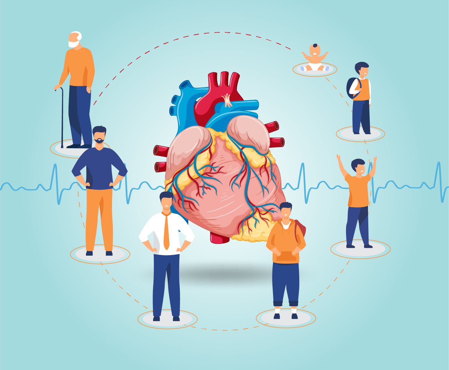
Pregnancy is a transformative and joyous experience in a woman’s life, but it also places significant physiological demands on the body. For women with pre-existing cardiovascular disease, pregnancy can present unique challenges and risks. Cardiovascular disease encompasses a range of conditions that affect the heart and blood vessels, including hypertension, congenital heart defects, valvular heart disease, and cardiomyopathy. Managing cardiovascular disease during pregnancy requires careful planning, monitoring, and collaboration between obstetricians and cardiologists to ensure the health of both the mother and the developing fetus.

Risk Factors and Preconception Care

Women with pre-existing cardiovascular disease who plan to become pregnant should receive specialized preconception care. This involves a thorough assessment of their cardiovascular health, including a review of their medical history, current medications, and cardiac function. Understanding the specific type and severity of cardiovascular disease is essential for risk assessment and management planning.

Risks for Mother

During pregnancy, the body undergoes significant changes to support fetal growth. Blood volume increases, heart rate rises, and the cardiovascular system works harder to meet the body’s increased oxygen and nutrient demands. For women with cardiovascular disease, these changes can aggravate their condition and lead to complications such as heart failure, arrhythmias, and thromboembolic events. Hypertension, in particular, can become more challenging to manage during pregnancy and is a significant risk factor for complications.

Risks for Fetus

The developing fetus relies on a well-functioning maternal cardiovascular system for oxygen and nutrient supply. Maternal cardiovascular disease can compromise blood flow to the placenta, potentially resulting in fetal growth restriction, preterm birth, or stillbirth. Additionally, certain medications used to manage cardiovascular conditions may have teratogenic effects on the developing fetus, highlighting the importance of careful medication management during pregnancy.

Management and Monitoring

Managing cardiovascular disease during pregnancy requires a multidisciplinary approach involving obstetricians, cardiologists, and other specialists as needed. Individualized treatment plans are crucial, taking into account the specific cardiovascular condition, its severity, and the patient’s overall health. Medications may need to be adjusted or changed to minimize potential harm to the fetus while ensuring maternal well-being.

Frequent monitoring is essential throughout pregnancy. This includes regular check-ups to assess blood pressure, cardiac function, and fetal growth. Advanced imaging techniques like echocardiography and Doppler ultrasound can provide valuable insights into maternal and fetal well-being.

Delivery Planning

The mode of delivery should also be carefully planned. In many cases, vaginal delivery is safe and appropriate, but a caesarean section may be recommended if there are concerns about the mother’s cardiovascular stability or the fetus’s well-being. The timing of delivery may also be adjusted to optimize outcomes for both mother and baby.

Postpartum Care

The challenges of managing cardiovascular disease do not end with delivery. The postpartum period is a critical time, as the body undergoes significant physiological changes once again. Close monitoring should continue, and adjustments to medications and treatment plans may be necessary.

Conclusion

Pregnancy with pre-existing cardiovascular disease requires meticulous planning, close monitoring, and a collaborative healthcare team. While it can be associated with increased risks for both the mother and the fetus, with proper management and care, many women with cardiovascular disease can have successful pregnancies and healthy babies. Education, counseling, and ongoing support are vital for empowering women with cardiovascular disease to make informed decisions about family planning and to navigate the complex journey of pregnancy with confidence.

Congenital Heart Disease in Children

Congenital heart disease (CHD) is a group of structural heart defects present at birth, affecting millions of children worldwide. These anomalies range from minor issues that may require minimal intervention to severe, life-threatening conditions. Understanding CHD, its causes, diagnosis, treatment, and the emotional toll it takes on families is crucial for providing comprehensive care and support.

Causes and Types of CHD

CHD originates during fetal development when the heart does not form correctly. While the exact causes remain unknown in many cases, genetic factors, maternal illnesses, and environmental factors may contribute. There are various types of CHD, classified into two main categories: cyanotic and acyanotic.

  • Cyanotic CHD: These defects cause decreased oxygen levels in the blood, resulting in bluish skin and lips. Examples include Tetralogy of Fallot and Transposition of the Great Arteries.
  • Acyanotic CHD: These defects do not lead to bluish discoloration but can still be serious. Examples include Ventricular Septal Defect (VSD) and Atrial Septal Defect (ASD).

Diagnosis and Screening

CHD is typically diagnosed during pregnancy through routine ultrasounds or shortly after birth when a newborn is screened for congenital anomalies. Early detection is crucial for prompt intervention and better outcomes.

Treatment and Management

The treatment approach for CHD varies depending on the type and severity of the defect. Some children may only require periodic monitoring, while others need surgical or interventional procedures. Advances in medical technology and surgical techniques have greatly improved the outlook for children with CHD.

  1. Medication: Some CHD cases can be managed with medication to control symptoms and improve heart function. For example, diuretics may help remove excess fluid buildup, while beta-blockers can regulate heart rate.
  2. Surgery: Surgical intervention is often necessary for complex CHD cases. Surgeons can repair heart defects, replace damaged valves, or even perform heart transplants when required.
  3. Interventional Procedures: Minimally invasive procedures like catheterization can be used to treat certain CHD cases. These procedures involve threading a catheter through blood vessels to repair or correct heart defects.

Psychosocial Impact

CHD not only affects the child but also places a significant emotional and financial burden on families. Parents often experience feelings of guilt, anxiety, and stress. Siblings may feel neglected, and the child with CHD may struggle with self-esteem and body image issues as they grow.

Supportive care, counseling, and access to support groups can help families navigate these challenges. A multidisciplinary approach involving pediatricians, cardiologists, social workers, and psychologists is essential to address both the medical and emotional aspects of CHD.

Long-term Outlook

Advancements in medical science have led to a brighter outlook for children with CHD. Many go on to lead healthy lives with appropriate care and monitoring. However, long-term follow-up is critical to identify and address any potential complications as they grow.

Conclusion

Congenital heart disease in children is a complex and challenging condition that requires comprehensive care and support. Early diagnosis and access to specialized medical care are vital for improving outcomes. Equally important is the emotional and psychosocial support provided to families as they navigate the journey of caring for a child with CHD. With continued research and advancements in medical technology, the future looks promising for children born with these heart defects, offering hope for healthier and happier lives.

Ventricular Tachycardia

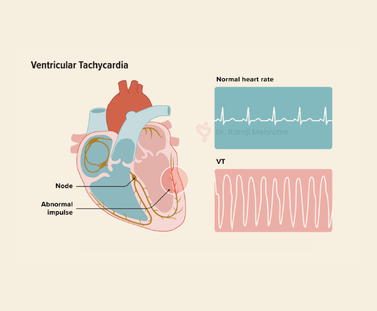
Ventricular tachycardia (VT) is a potentially life-threatening cardiac arrhythmia characterized by a rapid heartbeat originating in the ventricles, the lower chambers of the heart. In VT, the heart beats faster than the normal rate, usually over 100 beats per minute, and it can be sustained for longer periods, making it a serious medical condition that requires prompt attention and treatment.

Causes of Ventricular Tachycardia

VT typically results from abnormal electrical signals in the heart, often due to an underlying heart condition. Common causes include:

  1. Coronary Artery Disease (CAD): The most common cause of VT is CAD, which leads to insufficient blood flow to the heart muscle. This lack of oxygen can disrupt the heart’s electrical system, leading to arrhythmias like VT.
  2. Cardiomyopathy: Conditions like dilated or hypertrophic cardiomyopathy can cause changes in the heart’s structure and electrical system, increasing the risk of VT.
  3. Heart Attack: An acute myocardial infarction (heart attack) can scar heart tissue, creating areas where abnormal electrical impulses can arise.
  4. Valvular Heart Disease: Malfunctioning heart valves can affect blood flow, increasing the likelihood of arrhythmias, including VT.
  5. Electrolyte Imbalances: Abnormal levels of electrolytes (such as potassium, sodium, or calcium) can disrupt the heart’s electrical signals.

Symptoms

The symptoms of VT can vary in severity and may include:

  • Palpitations: A strong, rapid heartbeat is a hallmark symptom of VT.
  • Chest Pain: Some people may experience chest discomfort or pain during VT episodes.
  • Dizziness or Lightheadedness: Insufficient blood flow to the brain can lead to these sensations.
  • Shortness of Breath: Rapid heartbeats can reduce the heart’s efficiency, leading to breathlessness.
  • Loss of Consciousness: In severe cases, VT can cause fainting or even sudden cardiac arrest.

Diagnosis

A proper diagnosis of VT involves a combination of medical history, physical examination, and diagnostic tests, including:

  1. Electrocardiogram (ECG or EKG): This test records the heart’s electrical activity and can identify VT patterns.
  2. Holter Monitor: A portable ECG device worn for an extended period can capture intermittent VT episodes.
  3. Electrophysiology Study (EPS): Invasive testing, where thin wires are inserted into the heart, can pinpoint the location and cause of VT.
  4. Echocardiogram: This ultrasound imaging of the heart can reveal structural abnormalities.

Treatment Options

The management of VT depends on its cause, severity, and the patient’s overall health. Treatment options may include:

  1. Medications: Antiarrhythmic drugs can help control VT episodes by stabilizing the heart’s electrical activity.
  2. Implantable Cardioverter-Defibrillator (ICD): For those at high risk of life-threatening VT, an ICD is surgically implanted to monitor and correct abnormal rhythms by delivering shocks when needed.
  3. Catheter Ablation: This procedure involves the use of catheters to target and destroy the abnormal heart tissue responsible for VT.
  4. Lifestyle Changes: Managing underlying conditions like CAD or cardiomyopathy through lifestyle modifications, such as diet, exercise, and smoking cessation, can reduce the risk of VT.
  5. Emergency Care: If VT leads to cardiac arrest, immediate defibrillation is crucial to restore normal rhythm and save a life.

Conclusion

Ventricular tachycardia is a serious cardiac arrhythmia that requires careful evaluation and management. Early diagnosis and appropriate treatment are essential to reduce the risk of life-threatening complications. Patients with a history of heart disease or risk factors should consult with the doctors regularly for them to monitor and address any cardiac issues, ultimately improving their overall heart health and quality of life.

Silent heart attack: What are the risks?

A silent heart attack, medically termed a “silent myocardial infarction,” is a potentially dangerous event that often goes unnoticed due to its lack of typical symptoms. Unlike a traditional heart attack, which is accompanied by chest pain, shortness of breath, and discomfort in the arms, neck, jaw, or back, a silent heart attack occurs without these apparent signs. This makes it a significant concern, as individuals are less likely to seek immediate medical attention, leading to delayed diagnosis and treatment. Understanding the risks associated with silent heart attacks is crucial for prevention and proactive healthcare management.

Causes and Risks

Silent heart attacks share the same underlying cause as symptomatic heart attacks: a disruption of blood flow to a portion of the heart muscle due to a blocked or narrowed coronary artery. This interruption leads to the death of heart muscle cells, which can impair cardiac function and increase the risk of future heart-related complications. The factors that contribute to silent heart attacks are often the same as those for traditional heart attacks, including high blood pressure, high cholesterol, diabetes, obesity, smoking, and a sedentary lifestyle. However, certain demographic groups, such as older adults and individuals with diabetes, are more prone to experiencing silent heart attacks.

One of the major dangers of silent heart attacks is their gradual progression. Without the characteristic chest pain that typically prompts individuals to seek medical help, silent heart attacks can go undetected for weeks, months, or even years. During this time, untreated heart damage may accumulate, putting individuals at greater risk of heart failure, abnormal heart rhythms (arrhythmias), and subsequent heart attacks. Additionally, silent heart attacks are often identified incidentally when a person undergoes medical tests for unrelated issues, further highlighting the hidden nature of these events.

Preventive Measures

The lack of awareness surrounding silent heart attacks highlights the importance of preventive healthcare measures. Regular check-ups and screenings are essential, especially for individuals with risk factors. Blood pressure, cholesterol, and blood sugar levels should be closely monitored, and lifestyle modifications should be adopted to mitigate these risk factors. Adopting a heart-healthy diet rich in fruits, vegetables, whole grains, lean proteins, and healthy fats, along with engaging in regular physical activity, can significantly reduce the likelihood of both silent and symptomatic heart attacks.

Moreover, recognizing potential warning signs that might not be as dramatic as traditional symptoms is vital. Unexplained fatigue, mild discomfort in the chest, nausea, or discomfort in the upper abdomen, back, or jaw could all be indicative of a silent heart attack. Hence, they should not be ignored, especially if they persist or worsen over time.

Conclusion

Silent heart attacks pose a grave risk due to their inconspicuous nature and delayed diagnosis. The absence of classic symptoms can lead to untreated heart damage and increase the likelihood of complications. It is essential for individuals, particularly those with risk factors, to prioritize regular health check-ups, maintain a heart-healthy lifestyle, and be vigilant about any unusual or persistent symptoms. By taking proactive measures, individuals can mitigate the risks associated with silent heart attacks and promote overall heart health.

Different Types of Heart Disease

Heart disease, also known as cardiovascular disease, refers to a range of conditions that affect the heart and blood vessels. The heart is a vital organ that pumps blood throughout the body, and its proper functioning is essential for good health. However, various factors such as genetics, lifestyle choices, and other health conditions can contribute to the development of heart disease. In this blog we will discuss different types of heart diseases and how they affect the heart and blood vessels.

  • Coronary Artery Disease (CAD)

Coronary artery disease is a condition that occurs when the blood vessels that supply blood to the heart become narrow or blocked, causing a reduction in blood flow to the heart muscle especially when demand is high on exertion. This can lead to chest pain, angina, and heart attack. The primary cause of CAD is the build-up of plaque in the coronary arteries. Plaque is a mixture of cholesterol, fat, and other substances that can clog the arteries and make it difficult for blood to flow. The risk factors for CAD include high cholesterol, high blood pressure, smoking, obesity, and a family history of heart disease.

  • Heart Attack

A heart attack, also known as myocardial infarction, occurs when a blood clot or plaque ruptures in a coronary artery, blocking blood flow to a portion of the heart muscle. This can cause damage or death to the heart muscle, which can lead to serious health problems, including heart failure. The symptoms of a heart attack include chest pain, shortness of breath, sweating, and light headedness. Quick treatment with medications and procedures such as angioplasty or bypass surgery can help prevent further damage to the heart and reduce the risk of heart failure.

  • Heart Failure

During heart failure the heart is unable to pump sufficient blood to meet the body’s requirements. This can occur due to damage to the heart muscle from a heart attack, heart valve dysfunction, myopathy, certain infections, high blood pressure, or other health conditions. Heart failure can cause a number of symptoms, including fatigue, shortness of breath, and swelling in the legs, ankles, and feet. Treatment for heart failure includes medications, lifestyle changes, and in severe cases, surgery to implant a pacemaker or defibrillator, ventricular assist devices and heart transplant.

  • Arrhythmia

Arrhythmia is an irregular heartbeat that can occur due to a variety of factors, including heart disease, high blood pressure, and stress. The symptoms of arrhythmia include palpitations, dizziness, and shortness of breath. Treatment for arrhythmia may include medications, lifestyle changes, and procedures such as ablation or implantation of a pacemaker or defibrillator.

  • Valvular Heart Disease

Valvular heart disease is a condition that occurs when the valves in the heart that regulate blood flow become damaged or diseased. The most common types of valvular heart disease include aortic stenosis, in which the aortic valve becomes narrow, aortic regurgitation in which the valve leaks and mitral stenosis in which mitral valve becomes narrow and mitral regurgitation, in which the mitral valve leaks. The symptoms of valvular heart disease can include chest pain, shortness of breath, and fatigue. Treatment may include medications, lifestyle changes, or in severe cases, surgery to repair or replace the affected valve. In selected cases percutaneous valve replacement and repair is also possible nowadays.

  • Congenital Heart Disease

Congenital heart disease is a condition that is present at birth and affects the structure of the heart. This type of heart disease can be caused by genetic factors or environmental factors during pregnancy. The symptoms of congenital heart disease can vary depending on the specific condition, but may include shortness of breath, fatigue, and heart murmur. Treatment may include medications, lifestyle changes, or surgery to repair or replace the affected heart structures.

Conclusion

In conclusion, heart disease is a complex and often life-threatening condition that can take many forms. Understanding the different types of heart disease, their symptoms, and their causes, is an important step towards reducing the risk factors and improving the chances of maintaining good health. With timely treatment the patients can look forward to almost normal life in most cases.

What is Patent Ductus Arteriosus (PDA) Defect?

Patent ductus arteriosus is a very common heart condition found in new born babies. It occurs because the ductus arteriosus blood vessel doesn’t close properly after birth.

Babies born prematurely are more likely to be affected by PDA. The risk of PDA increases the earlier the baby is born. PDA affects 10% of babies born between 30 and 37 weeks of pregnancy, 80% of babies born between 25 and 28 weeks of pregnancy and 90% of babies born earlier than 24 weeks of pregnancy.

PDA is also more commonly seen in girls than in boys.

Cause of PDA

Every baby has a ductus arteriosus, which is a foetal artery connecting the aorta and the pulmonary artery. This opening usually narrows and closes within a few days after birth. If it continues to remain open it is known as patent ductus arteriosus (PDA).

While a small PDA may not generally cause any problems, a large PDA can lead to the flow of poorly oxygenated blood in the wrong direction. This can cause heart failure and other complications such as pulmonary hypertension and blood vessel damage.

Causes of PDA include genetic disorders or a family history of the condition. Babies with neonatal respiratory distress syndrome which is a breathing problem or those born to mothers who had rubella during pregnancy are also at risk of developing PDA.

Symptoms

PDA symptoms vary according to the size of the defect. Small PDAs may not have symptoms apart from a heart murmur and may go undetected until adulthood.

Large PDAs may cause symptoms such as:

  • Persistent rapid breathing.
  • Breathlessness
  • Sweating often
  • Fatigue
  • or tiredness
  • Feeding and eating problems
  • Poor weight gain
  • Rapid heart rate

It is important to consult a doctor immediately if any of these symptoms are noticed in the baby.

Diagnosis and Treatment

PDA in premature babies and in neonates may be the cause of respiratory compromise. Many a time’s medicine is effective. In cases where medicine has failed or cannot be given due to many reasons, surgery is required. 

The paediatric cardiologist may recommend various tests such as Chest X-ray, Electrocardiogram (ECG), Echocardiogram, etc to diagnose the PDA.

A small PDA may close on its own as the child grows and may not require treatment. Doctors will prescribe the medication as required.

Large PDAs may require treatment that includes cardiac catheterization or surgery. Catheterization is a minimally invasive procedure where the doctors use a flexible tube or catheter and insert a coil through it into the heart to close the PDA. In PDA surgery an incision is made to the side of the chest and the PDA is closed with stiches or a metal clip.

Life after PDA treatment

After receiving treatment for PDA, children can lead normal, healthy lives. Activity levels, appetite, and growth will return to normal within a short duration. Periodic check-ups with the paediatric cardiologist will ensure that there are no heart or lung problems.

What is Acute Coronary Syndrome?

Acute coronary syndrome (ACS) refers to conditions where the blood supply to the heart muscle is suddenly blocked or severely reduced. It is a life threatening condition affecting millions of people every year and requires prompt diagnosis and care. Unstable angina and heart attack both are types of ACS.

Causes

The build-up of plaque in the arteries can block the blood flow to the heart. Plaque is a fatty substance made up of cholesterol, fat and other substances.

Gradual build-up of plaque can cause an artery to become very narrow and later completely blocked. The plaque can rupture suddenly leading to the formation of a blood clot that narrows or blocks the artery.

Symptoms

Some signs and symptoms of ACS include:

  • Chest pain or severe discomfort
  • Pain spreading from the chest to other parts of the body
  • Shortness of breath
  • Sudden, heavy sweating
  • Nausea
  • Fast or irregular heartbeat

Symptoms may vary depending on the patient’s age, sex and other medical conditions.

Risk factors

Certain risk factors increase the probability of developing ACS. People over the age of 45, those who are obese, and those who smoke are at risk.

Similarly, other conditions like high blood cholesterol, hypertension, diabetes and family history of heart disease also cause a risk of ACS.

Types of ACS

ACS includes three types of coronary artery disease that can damage heart tissue. These are:

  • Unstable angina: It comprises sudden and unexpected chest pain and is a warning sign of a heart attack.
  • NSTEMI: A Non-ST-elevation myocardial infarction (NSTEMI) is a heart attack in which the coronary arteries aren’t fully blocked.
  • STEMI: An ST-elevation myocardial infarction (STEMI) is a severe heart attack that occurs when the blood flow to the heart is fully blocked.

Diagnosis and Treatment

There are a number of tests that doctors may recommend to diagnose ACS including:

  • Electrocardiogram (ECG) – It measures the heart’s electrical activity.
  • Blood test – Blood tests such as troponin blood test can detect the cause of chest pain and the risk of a heart attack.
  • Echocardiogram – This test uses sound waves to detect if heart has been damaged or has any other problems.

Treatment for ACS may comprise medicines, surgery, or other procedures to treat the symptoms and restore blood flow to the heart. Doctors may prescribe different medicines such as aspirin, beta blockers, blood thinners, clot dissolving drugs, angiotensin converting enzyme (ACE) inhibitors, or nitroglycerin based on the patient’s condition.

Angioplasty procedure is performed to open a clogged artery using a long, thin tube known as catheter. Bypass surgery is performed to route the blood around the blocked artery.

Prevention

According to Dr Ramji Mehrotra, the risk of ACS can be reduced to a great extent by adopting a healthy lifestyle.

This includes consuming a balanced diet with plenty of whole grains, vegetables and fruits while limiting foods with high cholesterol and saturated fats. It is also important to maintain optimum weight by exercising regularly and avoiding smoking and tobacco in any form as it can damage the heart.

It is also advised to get regular preventive health screenings done and manage health conditions such as cholesterol, blood pressure and diabetes under control.

An Overview of Breast Cancer

Breast cancer is the second most common cancer diagnosed in women. It occurs mostly in women, though men can also get breast cancer (less than 1% of cases). With growing awareness and advancement in diagnosis and treatment, breast cancer survival rates have increased and deaths due to the disease have decreased steadily.

Breast cancer can occur at any age, though it is mostly diagnosed in patients over the age of 50.

Spread of breast cancer

Breast cancer can start in one or both breasts when the cells begin to grow out of control. These cells divide more rapidly than healthy cells, forming a lump or mass. Most breast lumps are benign and not malignant (cancerous). However, it is very important to get any breast lump checked by a specialist.

Cells may spread through the breast to the lymph nodes or other parts of the body.

Symptoms

Different people may exhibit different symptoms of breast cancer. Some signs and symptoms of breast cancer include the following:

  • New lump in the breast or underarm
  • Change in the size or shape of the breast
  • Irritation or dimpling of the skin over the breast
  • Redness or flaky skin in the nipple area
  • Fluid discharge from the nipple
  • Pain in any area of the breast

A few people may not show any symptoms at all though they are affected by breast cancer.

Causes

Breast cancer generally begins with the cells in the milk-producing ducts. It may also begin in the glandular tissue called lobules or in other cells within the breast.

According to medical research, hormonal, lifestyle and environmental factors can all increase the risk of breast cancer. As a woman’s age increases, the risk of breast cancer also rises.

Family history and genetics also have a role in determining the risk of the disease. Around 5% to 10% of the cases are due to single abnormal genes. These can be detected by genetic testing.

Other causes of breast cancer include smoking and drinking alcohol. In addition, patients with prior radiation therapy or hormone replacement therapy are at greater risk of developing breast cancer.

Diagnosis of breast cancer

There are several tests that may be recommended by the doctor to check for breast abnormalities. These tests can include:

  • Mammogram: Mammogram is an X-ray picture of the breast that helps doctors in detecting early signs of breast cancer.
  • Ultrasonography: It helps in diagnosing breast lumps or abnormalities by using sound waves to take pictures of the tissues inside the breast.
  • Magnetic resonance imaging (MRI): MRI produces detailed images of the interna structure of the breast using magnets and radio waves.
  • Positron emission tomography (PET) scanning: During a PET scan, a special dye is injected into the veins, after which the image of the affected areas is taken by the scanner.

Treatment of breast cancer

Breast cancer can be treated using a number of treatment options such as surgery, chemotherapy, hormone therapy, immunotherapy, radiation therapy and targeted drug therapy. Doctors may recommend one or more of these options based on factors like location and size of the tumour, spread of cancer to the other parts of the body, results of the lab tests, etc.

Surgery involves removing the cancerous portion of the patient’s breast and an area of normal tissue surrounding the tumour. It can be of different types such as lumpectomy, mastectomy, sentinel node biopsy, etc.

Chemotherapy may be recommended before a lumpectomy to shrink the tumour and sometimes after surgery to kill any remaining cancer cells and reduce the risk of recurrence.

Hormone therapy is mostly used after surgery to reduce the risk of breast cancer recurrence, though it may also be used before surgery to shrink the tumour or to treat the cancer that has spread to other parts of the body.

Immunotherapy helps the patient’s immune system to target and attack breast cancer cells.

Radiation therapy is usually given after a lumpectomy or mastectomy to kill remaining cancer cells. Targeted drug therapy may be recommended by doctors in cases where breast cancer has spread to other areas of the body.

Impact of breast cancer on heart health

Breast cancer and heart disease can sometimes overlap in patients, and occurrence of one condition can lead to the development or recurrence of the other.

In some patients the radiation therapy for breast cancer may lead to issues such as blocked arteries, abnormal heart rhythms, etc. Similarly chemotherapy can sometimes weaken the heart and cause blood clots and other issues.

Medical research has also found that survivors of breast cancer, who are subsequently affected by cardiovascular disease, have an increased probability of cancer recurrence.

According to Dr Ramji Mehrotra, a few risk factors are common for both heart disease and breast cancer, and a heart-healthy lifestyle including proper diet and exercise must be adopted by women to minimise the chances of developing either disease.

Conclusion

Though breast cancer cannot be prevented totally, women can take certain precautions to reduce the risk of discovering it at an advanced stage.

All women over the age of 40 should get a mammogram screening done yearly. Doctors should be consulted with immediately whenever the women find any abnormality or changes in their breasts. Clinical breast exams can detect lumps that mammograms may not find.

Women with early-stage breast cancer can manage their condition successfully with treatment and live long, happy lives.

Women and Heart Disease

It is generally considered that heart disease affects men more than women. However, coronary heart disease is a leading cause of death for both men and women, though the symptoms may be different for both. According to research, cardiovascular disease accounts for 1 in every 3 deaths around the world.

Women and men experience heart disease differently due to the differences in the respective cardiovascular system. For example, women have smaller heart chambers and blood vessels than men. They have lesser number of red blood cells and cannot take in as much oxygen as men. Sudden drop in blood pressure and fainting are more likely in women.

Oestrogen and progesterone hormones are dominant in women, while in men it is the testosterone hormone. These also impact heart health differently.

Identification of heart disease in women

In women symptoms of cardiovascular disease are generally noticed much later than in men. The most common symptom is chest pain or angina which may feel like heaviness, pressure, aching, numbness, squeezing, etc. 

Other symptoms usually observed include fatigue, breathlessness, heart palpitations, nausea, sudden sweating, etc. Anxiety, loss of appetite, frequent indigestion, intense headache and discomfort in the jaws or teeth, are other warning signs of a possible heart attack.

Women are also at higher risk for having a silent heart attack, that is an attack without any visible symptoms.

Hence, it is very essential to get medical help whenever any unusual symptoms are noticed to prevent the chances of heart attack.

Risk factors for heart disease in women

While risk factors like high cholesterol and high blood pressure affect both men and women, women face certain unique risks that affect them more. These include the following:

  • Obesity: During menopause women face greater risk of obesity and gaining abdominal fat which puts them at higher risk of heart disease.
  • High cholesterol: In women aged 65 and above, low level of HDL or good cholesterol is closely linked with death, more than in men.
  • Diabetes: Women with diabetes have a much greater risk of developing cardiovascular disease than men with diabetes.
  • Hypertension: Hypertension or high blood pressure has been found to be linked more closely with heart attacks in women than in men.
  • Family history: Instances of early heart disease in the family cause greater risk to women than men.  

Reducing the risk of heart disease

According to Dr. Ramji Mehrotra, it is very important to adopt a healthy lifestyle in order to reduce the risk of heart disease. A healthy diet with whole grains, vegetables and fruits must be consumed. Saturated fats and high amounts of salt and sugar must be avoided to maintain good health.

Regular exercise is a must, especially for women who are overweight, in order to reduce the risk of heart disease. Stress must be kept under control with meditation and yoga, as high levels of stress have been found to cause microvascular disease.

In addition, other health conditions such as high blood pressure, diabetes and high cholesterol should be managed by regular consultations with the medical practitioners.

By following the above guidelines, heart disease can be prevented in women and they can lead healthy lives.

What is Coronary Artery Disease?

Coronary artery disease (CAD) or coronary heart disease is a common type of heart disease that occurs when the arteries become hardened and narrowed. This is caused due to the cholesterol and calcium deposit or plaque in the arteries.

Coronary artery disease begins due to a condition called atherosclerosis which occurs when cholesterol and calcium collects on the inner walls of the arteries. This build-up is known as plaque which can block blood flow due to the narrowing of the arteries. If the plaque bursts it can lead to a blood clot.

Symptoms of CAD

A patient having coronary artery disease can have chest pain, breathlessness, fatigue, nausea and due to the reduced blood flow to the heart. Chest discomfort or angina is accompanied by symptoms like numbness, heaviness, aching, squeezing, etc.

Complete blockage of blood flow can also lead to heart attack.

Risk Factors

In addition to high cholesterol, other causes of damage to coronary arteries include high blood pressure, diabetes, smoking, sedentary lifestyle, etc.

Obesity, stress, unhealthy diet, irregular sleep and alcohol usage are other risk factors for coronary artery disease.

Diagnosis of CAD

The doctor reviews the patient’s symptoms, risk factors and medical history before performing a physical examination. Diagnostic tests include:

  • Electrocardiograph test: This test can detect heart attack and heart rhythm issues by recording the heart’s electrical activity.
  • Exercise stress test: This is a treadmill test that can help suspect  coronary blockages
  • Pharmacologic stress test: This test can also help detect coronary blockages through medication given to increase heart rate. This is done in patients who cannot walk on treadmill.
  • Coronary calcium scan: This can identify the amount of calcium on the walls of the coronary arteries which can give an indication of atherosclerosis.
  • Echocardiogram: This test utilizes sound waves to measure the overall functioning of the heart.
  • Blood tests: These tests are done for factors affecting arteries like cholesterol, triglycerides, lipoprotein, glucose, etc.
  • Cardiac catheterization: In this test small tubes are inserted into the blood vessels to detect the presence of coronary artery disease and also evaluate the heart function.

Prevention of CAD

According to Dr. Ramji Mehrotra who is one of India’s leading cardiologist, coronary artery disease can be prevented by adopting a healthy lifestyle to improve heart health. These include stopping smoking and limiting alcohol use, consuming a heart-healthy diet, exercising and increasing activity levels, etc.

Blood pressure, diabetes and cholesterol should be controlled. Stress management is also essential to prevent coronary artery disease.

Posts navigation