Heart Disease and Pericardiocentesis

Heart disease is one of the leading causes of mortality and requires advanced medical interventions. Pericardiocentesis is a critical procedure in addressing complications related to pericardial effusion – a condition characterized by the accumulation of fluid in the pericardium, the protective sac around the heart. This procedure plays a vital role in both diagnosing and treating underlying heart conditions, alleviating symptoms, and preventing potentially life-threatening complications.

Causes

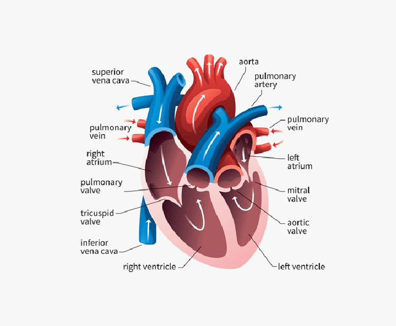
Pericardial effusion can result from various factors such as infections, inflammation, malignancies, trauma, or as a consequence of cardiac surgery. The accumulation of excess fluid within the pericardial space can exert pressure on the heart, impairing its ability to pump blood effectively. This can lead to symptoms like chest pain, shortness of breath, and even cardiac tamponade – a condition where the pressure from the fluid buildup compresses the heart chambers, compromising its function and circulation.

Procedure

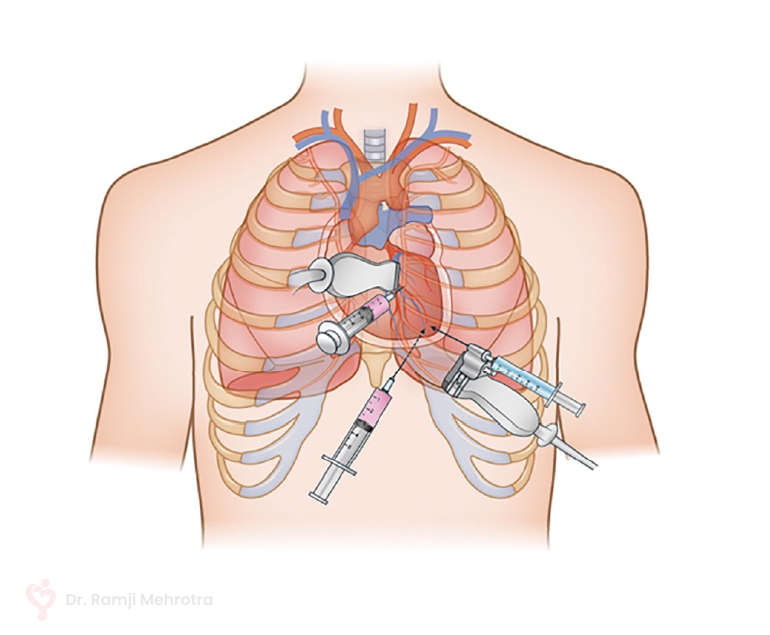
Pericardiocentesis involves the insertion of a needle or catheter into the pericardial space to drain the accumulated fluid. This procedure serves a dual purpose: providing symptomatic relief by relieving the pressure on the heart and facilitating diagnostic evaluation by analyzing the removed fluid for potential underlying causes.

The pericardiocentesis procedure is performed under imaging guidance, such as echocardiography or fluoroscopy, to ensure precision and minimize the risk of complications. The needle is carefully guided into the pericardial sac, and the fluid is aspirated. In some cases, a catheter may be left in place for continuous drainage if there is a significant fluid buildup or if the effusion is recurrent.

Benefits

Pericardiocentesis can provide rapid relief from the debilitating symptoms caused by pericardial effusion. By promptly reducing the pressure on the heart, patients often experience a marked improvement in their ability to breathe and reduced discomfort. Additionally, the removed fluid can be sent for analysis, which helps in diagnosing the underlying cause of the effusion. This diagnostic insight is crucial in guiding subsequent treatment decisions.

In some cases, pericardiocentesis may serve as a temporary measure while other interventions, such as medication, are initiated to address the underlying condition causing the effusion. However, in more severe cases where pericardial effusion poses an immediate threat to cardiac function, pericardiocentesis can be a life-saving intervention.

Conclusion

Pericardiocentesis plays a vital role in the management of heart disease, specifically in addressing the complications arising from pericardial effusion. This procedure offers both diagnostic and therapeutic benefits, helping in the swift mitigation of symptoms and potentially preventing life-threatening cardiac complications.

Rheumatic Heart Disease

Rheumatic heart disease (RHD) is a serious condition that affects the heart valves and is a result of complications from rheumatic fever. Although it is preventable and treatable, RHD continues to be a significant health burden in many developing countries, particularly among children and young adults.

Causes

RHD is caused by an abnormal immune response to untreated or inadequately treated streptococcal throat infections, mainly caused by group A streptococcus bacteria. When these infections are not promptly treated with antibiotics, the body’s immune system can mistakenly attack its own tissues, including the heart valves. Over time, this can lead to scarring, thickening, and deformation of the valves, which impairs their ability to function properly.

Symptoms

The symptoms of RHD can vary depending on the severity and location of valve damage. In the early stages, there may be no noticeable symptoms, but as the disease progresses, individuals may experience shortness of breath, chest pain, fatigue, palpitations, and swollen ankles or feet. In severe cases, heart failure and life-threatening complications, such as infective endocarditis or stroke, can occur.

Diagnosis & Treatment

Diagnosing RHD typically involves a combination of medical history assessment, physical examination, echocardiography (ultrasound of the heart), electrocardiogram (ECG), and other imaging tests. These tests help evaluate the structure and function of the heart, identify valve abnormalities, and determine the extent of damage.

Treatment for RHD aims to manage symptoms, prevent disease progression, and reduce the risk of complications. Antibiotic prophylaxis is crucial to prevent recurrent streptococcal infections, which can trigger disease exacerbation. Medications, such as diuretics, beta-blockers, and anticoagulants, may be prescribed to manage symptoms and reduce the risk of blood clots.

Prevention

Preventing RHD begins with prompt and appropriate treatment of streptococcal throat infections. Administering antibiotics, such as penicillin, to individuals with streptococcal infections can prevent the development of rheumatic fever, thereby reducing the risk of RHD. Regular follow-up and secondary prophylaxis, which involves long-term antibiotic treatment, are essential for individuals who had rheumatic fever to prevent disease recurrence and progression.

Conclusion

Rheumatic heart disease is a preventable and treatable condition that continues to be a significant health concern in many parts of the world. Prompt and appropriate treatment of streptococcal throat infections, along with regular follow-up and secondary prophylaxis, are vital for preventing the development and progression of RHD. By prioritizing prevention, early detection, and comprehensive management, the impact of rheumatic heart disease can be minimised.

What is an Aortic Stenosis Murmur?

Aortic stenosis murmur is a heart condition characterized by a narrowed opening in the aortic valve, which is the valve that controls blood flow from the heart to the rest of the body. This narrowing can cause a restriction in blood flow, leading to a number of symptoms and complications.

Symptoms of Aortic Stenosis Murmur

The most common symptom of aortic stenosis murmur is a heart murmur, which is a whooshing or whistling sound that can be heard through a stethoscope. This sound is caused by turbulent blood flow through the narrowed valve. Other symptoms can include chest pain, shortness of breath, fatigue, and fainting. In severe cases, aortic stenosis murmur can lead to heart failure, stroke, or even death.

Causes

There are several causes of aortic stenosis murmur. One of the most common causes is atherosclerosis, a condition in which plaque builds up in the arteries. This plaque can harden and narrow the aortic valve, making it difficult for blood to flow through. Other causes of aortic stenosis murmur include congenital heart defects, rheumatic fever, and aortic valve calcification.

Aortic stenosis murmur is usually diagnosed through a physical examination and listening for the heart murmur. Your healthcare provider may also use other diagnostic tests such as an echocardiogram, which uses sound waves to create a picture of the heart and its valves, or a cardiac catheterization, which involves threading a thin tube through an artery to the heart to measure pressure and blood flow.

Treatment

Treatment for aortic stenosis murmur depends on the severity of the condition. In mild cases, treatment may not be necessary and simply monitoring the condition may be sufficient. However, in more severe cases, treatment may be required to prevent complications and improve symptoms.

The most common treatment for aortic stenosis murmur is aortic valve replacement, which involves surgically removing the narrowed valve and replacing it with a new one. This can be done using either an artificial valve or a valve from a donor. Aortic valve replacement can significantly improve symptoms and reduce the risk of complications.

Another treatment option is aortic valve repair, which involves repairing the narrowed valve rather than replacing it. This can be done using techniques such as balloon valvuloplasty, which uses a balloon catheter to widen the valve, or valvulotomy, which involves cutting open the valve to widen it. Aortic valve repair can also significantly improve symptoms and reduce the risk of complications.

In some cases, a procedure called transcatheter aortic valve replacement (TAVR) may be used to replace the aortic valve. This procedure is less invasive than open heart surgery, and is done by threading a catheter through a blood vessel to the heart, and then deploying a new valve to replace the narrowed one.

In addition to these treatments, lifestyle changes can also help to improve symptoms and reduce the risk of complications. This can include quitting smoking, eating a healthy diet, getting regular exercise, and controlling blood pressure and cholesterol levels.

It is important to note that aortic stenosis murmur is a serious condition that requires prompt diagnosis and treatment. If you have symptoms of aortic stenosis murmur or are at risk of developing the condition, it is important to speak with your doctor about your options for diagnosis and treatment.

Coronary Microvascular Disease (CMD)

Coronary microvascular disease (CMD) is a condition that affects the small blood vessels in the heart. It is a common cause of chest pain and can also lead to heart failure. While CMD is often overlooked, it is a serious condition that can have significant consequences for people who have it.

How is CMD Caused?

CMD is caused by a blockage or damage to the small blood vessels in the heart, which can reduce blood flow to the heart muscle. This can cause chest pain, shortness of breath, and other symptoms. It can also lead to heart failure, in which the heart is unable to pump enough blood to meet the body’s needs.

CMD is often difficult to diagnose because the symptoms can be similar to those of other conditions such as angina or heart attack. In addition, traditional tests such as angiography may not be able to detect the blockages or damage in the small blood vessels. As a result, CMD may be misdiagnosed or go undiagnosed for some time.

Risk Factors

Risk factors for CMD include high blood pressure, high cholesterol, diabetes, and smoking. It is also more common in women and people with a family history of heart disease. These risk factors can damage the small blood vessels in the heart and lead to CMD.

Treatment of CMD

The good news is that CMD is a treatable condition. The first step in treatment is to manage any underlying risk factors such as high blood pressure or high cholesterol. This may involve lifestyle changes such as eating a healthy diet, getting regular exercise, and quitting smoking. In some cases, medications may be necessary to lower blood pressure and cholesterol levels and improve blood flow to the heart.

In addition to managing risk factors, it is important to address any symptoms of CMD. This may involve the use of medications to manage chest pain and improve blood flow to the heart. In some cases, procedures such as angioplasty or stenting may be necessary to open blocked blood vessels.

It is important to regularly consult with the specialist who can develop a treatment plan to manage CMD. This may include regular check-ups, blood pressure monitoring, and medication management. By taking steps to manage CMD, patients can reduce their risk of heart failure and other serious complications.

Conclusion

CMD is a serious condition that affects the small blood vessels in the heart. It can cause chest pain and lead to heart failure if left untreated. Risk factors for CMD include high blood pressure, high cholesterol, diabetes, and smoking. Treatment may involve lifestyle changes and medications, and in some cases, procedures to open blocked blood vessels.