CABG Procedure Overview

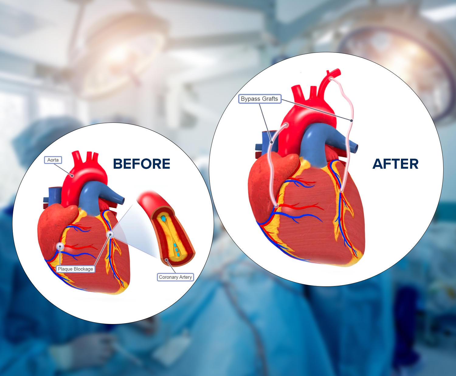
Coronary Artery Bypass Grafting (CABG) is a surgical procedure commonly performed to treat coronary artery disease (CAD), a condition in which the blood vessels supplying the heart muscle (coronary arteries) become narrowed or blocked. CABG is also known as heart bypass surgery. Here is an overview of the CABG procedure:

1. Preoperative Evaluation:

Medical History and Physical Examination: A physical examination is performed after assessing the patient’s medical history and overall health.

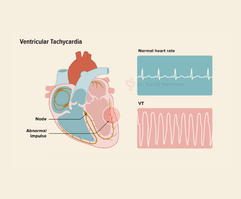
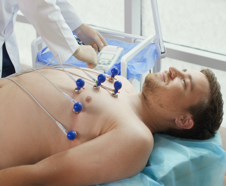
Diagnostic Tests: These may include an electrocardiogram (ECG or EKG), an echocardiogram, coronary angiography, and other imaging tests to evaluate the extent and location of coronary artery disease.

2. Anesthesia:

Pre-anesthetic Assessment: The anesthesiologist evaluates the patient’s overall health and determines the appropriate anesthesia plan.

General Anesthesia: During surgery, the patient is put under general anesthesia, which makes them unconscious and pain-free.

3. Harvesting of Grafts:

Graft Selection: Healthy blood vessels, often taken from the patient’s own body (autografts) or occasionally from a donor (allografts), are chosen for bypass grafts. Commonly used vessels include the internal mammary artery, radial artery, and saphenous vein.

4. Surgical Access:

Sternotomy or Thoracotomy: A sternotomy (cutting through the breastbone) is the most common approach, but in some cases, a thoracotomy (a small incision between the ribs) may be used.

5. Cardiopulmonary Bypass (if needed):

Connecting to a Heart-Lung Machine: The heart may be temporarily stopped, and blood is rerouted through a heart-lung machine, providing oxygen and circulation for the body while allowing the surgeon to work on a still heart.

6. Graft Placement:

Bypassing Blockages: The surgeon attaches one end of the graft to the aorta and the other end to a coronary artery beyond the blockage, creating a new pathway for blood flow.

7. Weaning off Bypass and Closing:

Restoration of Blood Flow: If a heart-lung machine is used, the patient is gradually weaned off it, and the heart resumes pumping.

Restoration of Blood Flow: If a heart-lung machine is used, the patient is gradually weaned off it, and the heart resumes pumping.

Closure: The chest has been closed, and the incision has been sutured.

8. Postoperative Care:

Monitoring: Initially, the patient is monitored closely in the intensive care unit (ICU) before being moved to a regular room.

Recovery: Rehabilitation and recovery involve medications, lifestyle changes, and follow-up care to ensure a smooth recovery.

9. Potential Risks and Complications:

Infection, bleeding, or blood clots: As with any surgery, there are potential risks, and complications can arise. The medical team closely monitors the patient to address any issues promptly.

According to Dr. Ramji MehrotaBest Heart Specialist Doctor in India, It’s important to note that the specifics of the procedure can vary based on individual patient characteristics and the surgeon’s preference. CABG is considered a major surgery, and the decision to undergo the procedure is made after a thorough assessment of the patient’s condition and the benefits of surgery. For personalized information, it’s best to consult healthcare professionals.

It’s important to note that the specifics of the procedure can vary based on individual patient characteristics and the surgeon’s preference. CABG is considered a major surgery, and the decision to undergo the procedure is made after a thorough assessment of the patient’s condition and the benefits of surgery. For personalized information, it’s best to consult healthcare professionals.

Ventricular Tachycardia

Ventricular tachycardia (VT) is a potentially life-threatening cardiac arrhythmia characterized by a rapid heartbeat originating in the ventricles, the lower chambers of the heart. In VT, the heart beats faster than the normal rate, usually over 100 beats per minute, and it can be sustained for longer periods, making it a serious medical condition that requires prompt attention and treatment.

Causes of Ventricular Tachycardia

VT typically results from abnormal electrical signals in the heart, often due to an underlying heart condition. Common causes include:

  1. Coronary Artery Disease (CAD): The most common cause of VT is CAD, which leads to insufficient blood flow to the heart muscle. This lack of oxygen can disrupt the heart’s electrical system, leading to arrhythmias like VT.
  2. Cardiomyopathy: Conditions like dilated or hypertrophic cardiomyopathy can cause changes in the heart’s structure and electrical system, increasing the risk of VT.
  3. Heart Attack: An acute myocardial infarction (heart attack) can scar heart tissue, creating areas where abnormal electrical impulses can arise.
  4. Valvular Heart Disease: Malfunctioning heart valves can affect blood flow, increasing the likelihood of arrhythmias, including VT.
  5. Electrolyte Imbalances: Abnormal levels of electrolytes (such as potassium, sodium, or calcium) can disrupt the heart’s electrical signals.

Symptoms

The symptoms of VT can vary in severity and may include:

  • Palpitations: A strong, rapid heartbeat is a hallmark symptom of VT.
  • Chest Pain: Some people may experience chest discomfort or pain during VT episodes.
  • Dizziness or Lightheadedness: Insufficient blood flow to the brain can lead to these sensations.
  • Shortness of Breath: Rapid heartbeats can reduce the heart’s efficiency, leading to breathlessness.
  • Loss of Consciousness: In severe cases, VT can cause fainting or even sudden cardiac arrest.

Diagnosis

A proper diagnosis of VT involves a combination of medical history, physical examination, and diagnostic tests, including:

  1. Electrocardiogram (ECG or EKG): This test records the heart’s electrical activity and can identify VT patterns.
  2. Holter Monitor: A portable ECG device worn for an extended period can capture intermittent VT episodes.
  3. Electrophysiology Study (EPS): Invasive testing, where thin wires are inserted into the heart, can pinpoint the location and cause of VT.
  4. Echocardiogram: This ultrasound imaging of the heart can reveal structural abnormalities.

Treatment Options

The management of VT depends on its cause, severity, and the patient’s overall health. Treatment options may include:

  1. Medications: Antiarrhythmic drugs can help control VT episodes by stabilizing the heart’s electrical activity.
  2. Implantable Cardioverter-Defibrillator (ICD): For those at high risk of life-threatening VT, an ICD is surgically implanted to monitor and correct abnormal rhythms by delivering shocks when needed.
  3. Catheter Ablation: This procedure involves the use of catheters to target and destroy the abnormal heart tissue responsible for VT.
  4. Lifestyle Changes: Managing underlying conditions like CAD or cardiomyopathy through lifestyle modifications, such as diet, exercise, and smoking cessation, can reduce the risk of VT.
  5. Emergency Care: If VT leads to cardiac arrest, immediate defibrillation is crucial to restore normal rhythm and save a life.

Conclusion

Ventricular tachycardia is a serious cardiac arrhythmia that requires careful evaluation and management. Early diagnosis and appropriate treatment are essential to reduce the risk of life-threatening complications. Patients with a history of heart disease or risk factors should consult with the doctors regularly for them to monitor and address any cardiac issues, ultimately improving their overall heart health and quality of life.

Dobutamine Stress Echocardiogram

Dobutamine Stress Echocardiogram, also known as DSE, is a non-invasive diagnostic test used to evaluate the function and blood flow of the heart. This test is commonly performed to detect and assess the severity of coronary artery disease or other conditions that may affect the heart’s ability to pump blood efficiently.

During a DSE, the patient is given a medication called dobutamine, which is a synthetic version of the hormone dopamine. This medication stimulates the heart, causing it to beat faster and harder, similar to what happens during exercise. As a result, the heart’s blood flow and function can be evaluated under conditions of increased stress.

An echocardiogram machine is used to generate images of the heart, while the dobutamine is administered through an intravenous (IV) line. The dose of dobutamine is gradually increased over several stages, while the heart is monitored using electrocardiogram (ECG) and echocardiogram (ultrasound) equipment.

The test is divided into several stages, and during each stage, the patient’s heart rate and blood pressure are monitored to evaluate the heart’s response to the medication. The stages are typically categorized based on the level of stress placed on the heart, ranging from rest to peak exertion. The test usually takes about 30 to 60 minutes to complete, depending on the patient’s response to the medication.

One of the main advantages of a DSE over other types of stress tests is that it does not require physical exertion, making it a safer option for patients who may not be able to exercise due to other medical conditions. Additionally, a DSE provides a more comprehensive evaluation of the heart’s function than other non-invasive tests, such as a resting echocardiogram or stress electrocardiogram.

The DSE test is commonly used to diagnose and assess the severity of coronary artery disease (CAD), which occurs when the arteries that supply blood to the heart become narrowed or blocked. CAD can lead to chest pain, heart attack, or other serious complications if left untreated. During a DSE, areas of the heart that are not receiving enough blood flow due to narrowed or blocked arteries can be identified, allowing doctors to determine the best treatment options.

In addition to CAD, a DSE can also be used to diagnose and evaluate other heart conditions, such as heart valve disease, cardiomyopathy, or pulmonary hypertension. By evaluating the heart’s function under conditions of increased stress, doctors can better understand how these conditions are affecting the heart’s ability to pump blood efficiently.

In conclusion, a Dobutamine Stress Echocardiogram is a safe and effective non-invasive test used to evaluate the function and blood flow of the heart. It is a valuable tool in the diagnosis and management of various heart conditions, including coronary artery disease and heart valve disease. By providing a comprehensive evaluation of the heart’s function under conditions of increased stress, doctors can better understand how these conditions are affecting the heart and develop an appropriate treatment plan.

What is Cardiac Catheterization?

Cardiac catheterization, also known as coronary angiography, is a medical procedure that allows doctors to examine the heart and blood vessels to identify any problems. It is a minimally invasive procedure that involves inserting a thin, flexible tube called a catheter into a blood vessel, typically in the groin or arm, and guiding it through the blood vessels to the heart.

The catheterization procedure is performed in a cardiac catheterization laboratory (also known as a cath lab) by a cardiologist. The procedure is typically done under local anaesthesia, which numbs the area where the catheter will be inserted.

Procedure

During the procedure, the cardiologist will make a small incision in the skin and insert a hollow sheath into the blood vessel. The catheter is then inserted through the sheath and guided to the heart using x-ray guidance. Once the catheter is in place, the cardiologist can inject a contrast dye into the bloodstream, which makes the heart and blood vessels visible on x-ray.

The contrast dye allows the cardiologist to examine the heart and blood vessels for any abnormalities or blockages that may be present. If a blockage is found, the cardiologist can perform a procedure called angioplasty to open the blocked artery and restore blood flow to the heart.

Diagnostic Tool

Cardiac catheterization is a valuable diagnostic tool that can provide important information about the heart and blood vessels. It is typically used to diagnose and evaluate a range of heart conditions, including coronary artery disease, heart valve problems, congenital heart defects, and heart failure.

In addition to providing diagnostic information, cardiac catheterization can also be used to treat certain heart conditions. For example, angioplasty can be used to open blocked arteries and restore blood flow to the heart. Other procedures, such as stent placement and balloon valvuloplasty, can also be performed during cardiac catheterization.

Risks

While cardiac catheterization is generally considered a safe procedure, there are some risks associated with it. These include bleeding, infection, damage to the blood vessels, and allergic reactions to the contrast dye. However, these risks are generally low and can be minimized with proper preparation and monitoring.

Tests before the procedure

Before undergoing cardiac catheterization, patients will typically undergo a series of tests to evaluate their heart health and determine if they are a good candidate for the procedure. These tests may include an electrocardiogram (ECG), echocardiogram, stress test, and blood tests.

Patients should also inform their doctor of any medications they are taking, as some medications may need to be temporarily stopped prior to the procedure. Patients should also avoid eating or drinking anything for several hours before the procedure.

Conclusion

Overall, cardiac catheterization is a valuable tool for diagnosing and treating a range of heart conditions. While there are some risks associated with the procedure, these risks are generally low and can be minimized with proper preparation and monitoring. Patients should talk to their doctor about whether cardiac catheterization is a good option for them and what they can expect during and after the procedure.

Complete Atrioventricular Canal Defect (CAVC)

Complete atrioventricular canal defect (CAVC) is a rare congenital heart condition that occurs when there is a hole in the centre of the heart that affects the atria and ventricles. This condition is also known as an atrioventricular septal defect (AVSD), and it can cause a range of serious health problems if left untreated.

In a normal heart, there is a wall (septum) between the two atria and another wall between the two ventricles. These walls prevent the mixing of oxygen-rich and oxygen-poor blood. In CAVC, these walls are missing, and a large hole exists between the atria and ventricles. As a result, oxygen-rich blood mixes with oxygen-poor blood, leading to reduced oxygen levels in the body and heart failure over time.

Diagnosis of CAVC

CAVC is typically diagnosed in infancy or early childhood. Infants with CAVC may experience symptoms such as difficulty breathing, rapid breathing, and feeding difficulties. In some cases, babies with CAVC may also have low oxygen levels, which can cause a bluish tint to the skin (cyanosis).

The diagnosis of CAVC is usually confirmed through diagnostic tests such as an echocardiogram, which uses ultrasound waves to create images of the heart. Other tests that may be performed include an electrocardiogram (ECG) to evaluate the heart’s electrical activity and a chest X-ray to check for signs of heart enlargement.

Treatment

Treatment for CAVC usually involves surgery to close the hole in the heart and reconstruct the affected valves. This type of surgery is typically done in early infancy or early childhood, depending on the severity of the condition. In some cases, medication may be prescribed to manage symptoms such as heart failure or to prevent complications such as infection.

Long-term outlook

The long-term outlook for children with CAVC depends on the severity of the defect and the timing of treatment. In general, children who receive timely and appropriate treatment have a good prognosis and can live normal, healthy lives. However, in severe cases, CAVC can lead to heart failure, pulmonary hypertension, and other serious complications.

In addition to medical treatment, children with CAVC may require ongoing monitoring and care throughout their lives. This may include regular check-ups with a pediatric cardiologist, medication management, and follow-up diagnostic tests such as echocardiograms.

Conclusion

Complete atrioventricular canal defect is a rare but serious congenital heart condition that affects the atria and ventricles of the heart. Early diagnosis and appropriate treatment are crucial for managing the condition and preventing complications. If you suspect your child may have CAVC, it is important to seek medical attention promptly to ensure the best possible outcome.