Cardiovascular Disease and Pregnancy

Pregnancy is a transformative and joyous experience in a woman’s life, but it also places significant physiological demands on the body. For women with pre-existing cardiovascular disease, pregnancy can present unique challenges and risks. Cardiovascular disease encompasses a range of conditions that affect the heart and blood vessels, including hypertension, congenital heart defects, valvular heart disease, and cardiomyopathy. Managing cardiovascular disease during pregnancy requires careful planning, monitoring, and collaboration between obstetricians and cardiologists to ensure the health of both the mother and the developing fetus.

Risk Factors and Preconception Care

Women with pre-existing cardiovascular disease who plan to become pregnant should receive specialized preconception care. This involves a thorough assessment of their cardiovascular health, including a review of their medical history, current medications, and cardiac function. Understanding the specific type and severity of cardiovascular disease is essential for risk assessment and management planning.

Risks for Mother

During pregnancy, the body undergoes significant changes to support fetal growth. Blood volume increases, heart rate rises, and the cardiovascular system works harder to meet the body’s increased oxygen and nutrient demands. For women with cardiovascular disease, these changes can aggravate their condition and lead to complications such as heart failure, arrhythmias, and thromboembolic events. Hypertension, in particular, can become more challenging to manage during pregnancy and is a significant risk factor for complications.

Risks for Fetus

The developing fetus relies on a well-functioning maternal cardiovascular system for oxygen and nutrient supply. Maternal cardiovascular disease can compromise blood flow to the placenta, potentially resulting in fetal growth restriction, preterm birth, or stillbirth. Additionally, certain medications used to manage cardiovascular conditions may have teratogenic effects on the developing fetus, highlighting the importance of careful medication management during pregnancy.

Management and Monitoring

Managing cardiovascular disease during pregnancy requires a multidisciplinary approach involving obstetricians, cardiologists, and other specialists as needed. Individualized treatment plans are crucial, taking into account the specific cardiovascular condition, its severity, and the patient’s overall health. Medications may need to be adjusted or changed to minimize potential harm to the fetus while ensuring maternal well-being.

Frequent monitoring is essential throughout pregnancy. This includes regular check-ups to assess blood pressure, cardiac function, and fetal growth. Advanced imaging techniques like echocardiography and Doppler ultrasound can provide valuable insights into maternal and fetal well-being.

Delivery Planning

The mode of delivery should also be carefully planned. In many cases, vaginal delivery is safe and appropriate, but a caesarean section may be recommended if there are concerns about the mother’s cardiovascular stability or the fetus’s well-being. The timing of delivery may also be adjusted to optimize outcomes for both mother and baby.

Postpartum Care

The challenges of managing cardiovascular disease do not end with delivery. The postpartum period is a critical time, as the body undergoes significant physiological changes once again. Close monitoring should continue, and adjustments to medications and treatment plans may be necessary.

Conclusion

Pregnancy with pre-existing cardiovascular disease requires meticulous planning, close monitoring, and a collaborative healthcare team. While it can be associated with increased risks for both the mother and the fetus, with proper management and care, many women with cardiovascular disease can have successful pregnancies and healthy babies. Education, counseling, and ongoing support are vital for empowering women with cardiovascular disease to make informed decisions about family planning and to navigate the complex journey of pregnancy with confidence.

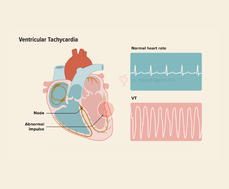
Ventricular Tachycardia

Ventricular tachycardia (VT) is a potentially life-threatening cardiac arrhythmia characterized by a rapid heartbeat originating in the ventricles, the lower chambers of the heart. In VT, the heart beats faster than the normal rate, usually over 100 beats per minute, and it can be sustained for longer periods, making it a serious medical condition that requires prompt attention and treatment.

Causes of Ventricular Tachycardia

VT typically results from abnormal electrical signals in the heart, often due to an underlying heart condition. Common causes include:

  1. Coronary Artery Disease (CAD): The most common cause of VT is CAD, which leads to insufficient blood flow to the heart muscle. This lack of oxygen can disrupt the heart’s electrical system, leading to arrhythmias like VT.
  2. Cardiomyopathy: Conditions like dilated or hypertrophic cardiomyopathy can cause changes in the heart’s structure and electrical system, increasing the risk of VT.
  3. Heart Attack: An acute myocardial infarction (heart attack) can scar heart tissue, creating areas where abnormal electrical impulses can arise.
  4. Valvular Heart Disease: Malfunctioning heart valves can affect blood flow, increasing the likelihood of arrhythmias, including VT.
  5. Electrolyte Imbalances: Abnormal levels of electrolytes (such as potassium, sodium, or calcium) can disrupt the heart’s electrical signals.

Symptoms

The symptoms of VT can vary in severity and may include:

  • Palpitations: A strong, rapid heartbeat is a hallmark symptom of VT.
  • Chest Pain: Some people may experience chest discomfort or pain during VT episodes.
  • Dizziness or Lightheadedness: Insufficient blood flow to the brain can lead to these sensations.
  • Shortness of Breath: Rapid heartbeats can reduce the heart’s efficiency, leading to breathlessness.
  • Loss of Consciousness: In severe cases, VT can cause fainting or even sudden cardiac arrest.

Diagnosis

A proper diagnosis of VT involves a combination of medical history, physical examination, and diagnostic tests, including:

  1. Electrocardiogram (ECG or EKG): This test records the heart’s electrical activity and can identify VT patterns.
  2. Holter Monitor: A portable ECG device worn for an extended period can capture intermittent VT episodes.
  3. Electrophysiology Study (EPS): Invasive testing, where thin wires are inserted into the heart, can pinpoint the location and cause of VT.
  4. Echocardiogram: This ultrasound imaging of the heart can reveal structural abnormalities.

Treatment Options

The management of VT depends on its cause, severity, and the patient’s overall health. Treatment options may include:

  1. Medications: Antiarrhythmic drugs can help control VT episodes by stabilizing the heart’s electrical activity.
  2. Implantable Cardioverter-Defibrillator (ICD): For those at high risk of life-threatening VT, an ICD is surgically implanted to monitor and correct abnormal rhythms by delivering shocks when needed.
  3. Catheter Ablation: This procedure involves the use of catheters to target and destroy the abnormal heart tissue responsible for VT.
  4. Lifestyle Changes: Managing underlying conditions like CAD or cardiomyopathy through lifestyle modifications, such as diet, exercise, and smoking cessation, can reduce the risk of VT.
  5. Emergency Care: If VT leads to cardiac arrest, immediate defibrillation is crucial to restore normal rhythm and save a life.

Conclusion

Ventricular tachycardia is a serious cardiac arrhythmia that requires careful evaluation and management. Early diagnosis and appropriate treatment are essential to reduce the risk of life-threatening complications. Patients with a history of heart disease or risk factors should consult with the doctors regularly for them to monitor and address any cardiac issues, ultimately improving their overall heart health and quality of life.

Heart Disease and Pericardiocentesis

Heart disease is one of the leading causes of mortality and requires advanced medical interventions. Pericardiocentesis is a critical procedure in addressing complications related to pericardial effusion – a condition characterized by the accumulation of fluid in the pericardium, the protective sac around the heart. This procedure plays a vital role in both diagnosing and treating underlying heart conditions, alleviating symptoms, and preventing potentially life-threatening complications.

Causes

Pericardial effusion can result from various factors such as infections, inflammation, malignancies, trauma, or as a consequence of cardiac surgery. The accumulation of excess fluid within the pericardial space can exert pressure on the heart, impairing its ability to pump blood effectively. This can lead to symptoms like chest pain, shortness of breath, and even cardiac tamponade – a condition where the pressure from the fluid buildup compresses the heart chambers, compromising its function and circulation.

Procedure

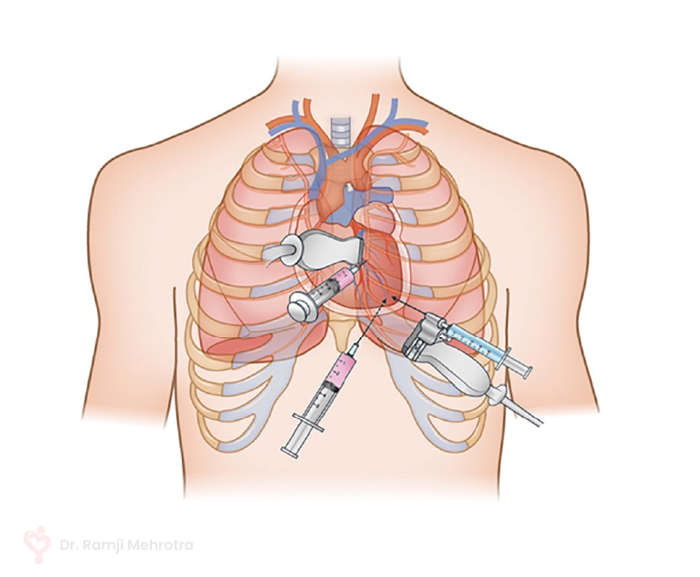
Pericardiocentesis involves the insertion of a needle or catheter into the pericardial space to drain the accumulated fluid. This procedure serves a dual purpose: providing symptomatic relief by relieving the pressure on the heart and facilitating diagnostic evaluation by analyzing the removed fluid for potential underlying causes.

The pericardiocentesis procedure is performed under imaging guidance, such as echocardiography or fluoroscopy, to ensure precision and minimize the risk of complications. The needle is carefully guided into the pericardial sac, and the fluid is aspirated. In some cases, a catheter may be left in place for continuous drainage if there is a significant fluid buildup or if the effusion is recurrent.

Benefits

Pericardiocentesis can provide rapid relief from the debilitating symptoms caused by pericardial effusion. By promptly reducing the pressure on the heart, patients often experience a marked improvement in their ability to breathe and reduced discomfort. Additionally, the removed fluid can be sent for analysis, which helps in diagnosing the underlying cause of the effusion. This diagnostic insight is crucial in guiding subsequent treatment decisions.

In some cases, pericardiocentesis may serve as a temporary measure while other interventions, such as medication, are initiated to address the underlying condition causing the effusion. However, in more severe cases where pericardial effusion poses an immediate threat to cardiac function, pericardiocentesis can be a life-saving intervention.

Conclusion

Pericardiocentesis plays a vital role in the management of heart disease, specifically in addressing the complications arising from pericardial effusion. This procedure offers both diagnostic and therapeutic benefits, helping in the swift mitigation of symptoms and potentially preventing life-threatening cardiac complications.

Tricuspid Valve Disease

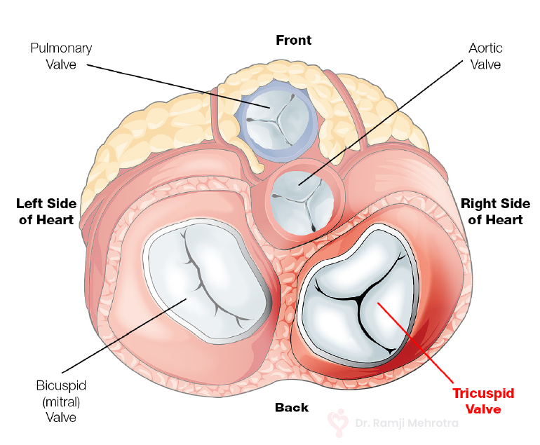
The tricuspid valve is an essential component of the heart and regulates blood flow between the right atrium and right ventricle. Tricuspid valve disease is a medical condition that affects the functionality of this valve, potentially leading to significant cardiovascular complications.

Categories

Tricuspid valve disease can be categorized into two main types: tricuspid regurgitation and tricuspid stenosis. Tricuspid regurgitation occurs when the valve doesn’t close properly, allowing blood to flow back into the right atrium during the ventricular contraction. This puts an extra strain on the heart and can lead to symptoms like fatigue, swelling in the legs and abdomen, and an enlarged liver.

On the other hand, tricuspid stenosis involves a narrowing of the valve, which restricts blood flow from the right atrium to the right ventricle. This condition can result in symptoms such as fatigue, palpitations, and fluid retention.

Causes

The causes of tricuspid valve disease can vary. It may be a result of congenital defects, where the valve doesn’t form properly during foetal development. Acquired tricuspid valve disease is often associated with other heart conditions, such as rheumatic fever, infective endocarditis, or pulmonary hypertension. These underlying conditions can damage the valve’s structure and function over time.

Diagnosis

Diagnosing tricuspid valve disease typically involves a combination of medical history assessment, physical examination, and various imaging techniques. Echocardiography is commonly used to visualize the valve’s structure and assess blood flow. Electrocardiograms (ECGs) and chest X-rays can help evaluate the heart’s electrical activity and overall condition.

Treatment

Treatment options for tricuspid valve disease depend on the severity of the condition and the associated symptoms. In mild cases, close monitoring and lifestyle modifications, such as reducing sodium intake and managing fluid retention, may be sufficient. For more severe cases, medical interventions or surgical procedures might be necessary.

Medical management often includes medications to alleviate symptoms and manage underlying conditions. Surgical interventions can range from minimally invasive techniques to open-heart surgery, depending on the complexity of the disease and the patient’s overall health.

Conclusion

Tricuspid valve disease is a complex cardiovascular condition that can have significant implications for a person’s health. Early detection and appropriate management are crucial to prevent the progression of the disease and the development of potentially life-threatening complications.

Hypertrophic Cardiomyopathy

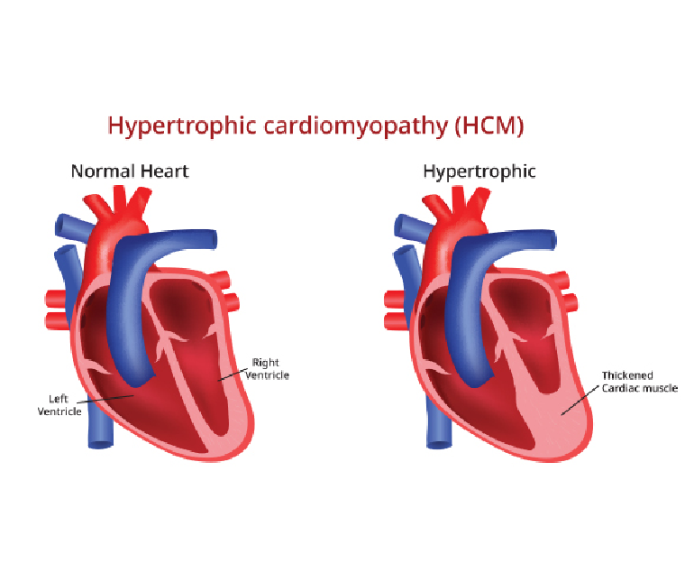
Hypertrophic cardiomyopathy (HOCM) is a complex and potentially life-threatening heart condition characterized by abnormal thickening (hypertrophy) of the heart muscle. It is one of the most common genetic heart disorders, affecting people of all ages and backgrounds.

Causes

Hypertrophic cardiomyopathy is primarily caused by mutations in genes responsible for the structure and function of the heart muscle. These genetic mutations lead to abnormal growth and arrangement of the cardiac muscle cells, resulting in the characteristic thickening of the heart muscle.

HOCM is usually inherited in an autosomal dominant pattern, meaning that a child has a 50% chance of inheriting the condition if one of the parents is affected. However, spontaneous gene mutations can also occur, leading to the development of HOCM in individuals without a family history of the condition.

Symptoms

The symptoms of hypertrophic cardiomyopathy can vary widely among individuals. Some may remain asymptomatic throughout their lives, while others may experience severe symptoms. Common symptoms include shortness of breath, chest pain, heart palpitations, fatigue, dizziness, fainting, and lightheadedness. These symptoms can worsen during physical exertion or strenuous activities and may be indicative of an increased risk of sudden cardiac arrest, especially in young athletes.

Diagnosis

The diagnosis of hypertrophic cardiomyopathy involves a comprehensive evaluation of the patient’s medical history, a physical examination, and a variety of diagnostic tests. These tests may include electrocardiography (ECG), echocardiography, cardiac MRI, exercise stress tests, and genetic testing. ECG can help identify abnormal electrical patterns in the heart, while echocardiography and cardiac MRI provide detailed images of the heart’s structure and function. Genetic testing is performed to identify specific gene mutations associated with HOCM.

Treatment

The management of hypertrophic cardiomyopathy focuses on relieving symptoms, preventing complications, and reducing the risk of sudden cardiac arrest. Treatment plans are tailored to individual patients based on their symptoms, risk factors, and disease progression. Medications such as beta-blockers, calcium channel blockers, and anti-arrhythmic drugs are commonly prescribed to control heart rate, rhythm, and blood pressure.

In some cases, surgical procedures like septal myectomy or alcohol septal ablation may be recommended to relieve obstruction of blood flow from the heart. Implantable cardioverter-defibrillators (ICDs) may also be used in high-risk patients to prevent sudden cardiac arrest.

Prognosis

The prognosis for individuals with hypertrophic cardiomyopathy varies depending on the severity of the disease, the presence of complications, and the effectiveness of treatment. Many people with HOCM lead normal lives with minimal symptoms and a low risk of complications. Regular monitoring, adherence to treatment plans, and lifestyle modifications, such as avoiding strenuous activities, can help improve outcomes and reduce the risk of complications.

Conclusion

Hypertrophic cardiomyopathy is a complex heart condition characterized by abnormal thickening of the heart muscle. It is primarily caused by genetic mutations and can present with a range of symptoms and complications. Early diagnosis, thorough evaluation, and personalized treatment plans are essential for managing HOCM effectively. With appropriate medical care and lifestyle modifications, individuals with hypertrophic cardiomyopathy can lead fulfilling lives while minimizing the risks associated with the condition.

Cardiovascular Conditions in Older Patients

Cardiovascular conditions in older patients pose significant challenges due to the natural aging process and the increased prevalence of risk factors associated with advanced age. As individuals grow older, their cardiovascular system undergoes changes that can increase the likelihood of developing various heart-related disorders.

Some of the common cardiovascular conditions seen in older patients include:

Coronary Artery Disease (CAD)

One of the most prevalent cardiovascular conditions in older adults is coronary artery disease (CAD). CAD occurs when the arteries that supply blood to the heart become narrowed or blocked due to atherosclerosis, the build-up of plaque. Older adults are more at risk to CAD due to factors such as prolonged exposure to risk factors like high blood pressure, high cholesterol, and smoking.

Diagnosis of CAD in older patients may be challenging because symptoms can be masked by other age-related conditions. Doctors employ diagnostic tools such as stress tests and cardiac imaging to confirm the diagnosis. Treatment typically involves a combination of lifestyle modifications, medication management, and, in severe cases, invasive interventions like angioplasty or bypass surgery.

Heart failure

Heart failure is another common cardiovascular condition in older patients. It occurs when the heart is unable to pump blood efficiently, leading to symptoms such as shortness of breath, fatigue, and fluid retention. Age-related changes in the heart, such as reduced elasticity and stiffening of the cardiac muscles, increase the risk of heart failure.

Accurate diagnosis of heart failure requires a comprehensive assessment of symptoms, physical examination, and diagnostic tests like echocardiography. Treatment involves a combination of medications to manage symptoms and improve heart function, lifestyle modifications like fluid and salt restriction, and, in some cases, devices like pacemakers or implantable defibrillators.

Arrhythmias

Arrhythmias, including atrial fibrillation (AF), are commonly encountered in older adults. AF, characterized by irregular and rapid heartbeat, is associated with an increased risk of stroke, heart failure, and other cardiovascular complications.

Advanced age is a significant risk factor for developing AF due to changes in the electrical conduction system of the heart. Diagnosis involves an electrocardiogram (ECG) and may require additional monitoring methods like Holter monitoring or event recorders. Management of AF in older patients aims to control heart rate, prevent blood clots with anticoagulant therapy, and restore normal heart rhythm when possible.

Hypertension

Hypertension, or high blood pressure, is highly prevalent in older adults and is a major risk factor for cardiovascular disease. Age-related changes in blood vessels, hormonal factors, and lifestyle habits contribute to the development of hypertension.

Effective management of hypertension in older patients involves lifestyle modifications, such as a heart-healthy diet and regular exercise, along with medications to achieve blood pressure targets. Close monitoring and regular follow-up are essential to ensure blood pressure control and prevent complications.

Conclusion

Cardiovascular conditions in older patients require a comprehensive and individualized approach to diagnosis and treatment. Age-related changes in the cardiovascular system increase the vulnerability of older adults to conditions like coronary artery disease, heart failure, arrhythmias, and hypertension. Addressing modifiable risk factors and promoting healthy lifestyle habits are essential for preventing and managing cardiovascular conditions in older patients.