Complete Atrioventricular Canal Defect (CAVC)

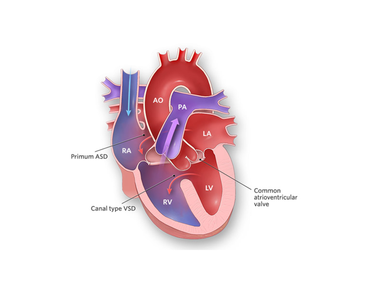
Complete atrioventricular canal defect (CAVC) is a rare congenital heart condition that occurs when there is a hole in the centre of the heart that affects the atria and ventricles. This condition is also known as an atrioventricular septal defect (AVSD), and it can cause a range of serious health problems if left untreated.

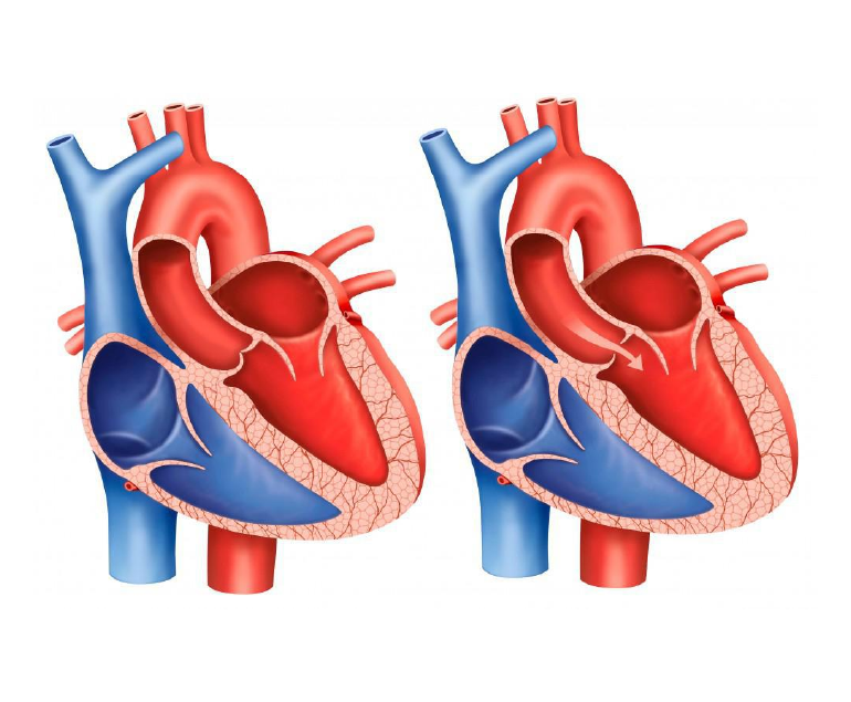
In a normal heart, there is a wall (septum) between the two atria and another wall between the two ventricles. These walls prevent the mixing of oxygen-rich and oxygen-poor blood. In CAVC, these walls are missing, and a large hole exists between the atria and ventricles. As a result, oxygen-rich blood mixes with oxygen-poor blood, leading to reduced oxygen levels in the body and heart failure over time.

Diagnosis of CAVC

CAVC is typically diagnosed in infancy or early childhood. Infants with CAVC may experience symptoms such as difficulty breathing, rapid breathing, and feeding difficulties. In some cases, babies with CAVC may also have low oxygen levels, which can cause a bluish tint to the skin (cyanosis).

The diagnosis of CAVC is usually confirmed through diagnostic tests such as an echocardiogram, which uses ultrasound waves to create images of the heart. Other tests that may be performed include an electrocardiogram (ECG) to evaluate the heart’s electrical activity and a chest X-ray to check for signs of heart enlargement.

Treatment

Treatment for CAVC usually involves surgery to close the hole in the heart and reconstruct the affected valves. This type of surgery is typically done in early infancy or early childhood, depending on the severity of the condition. In some cases, medication may be prescribed to manage symptoms such as heart failure or to prevent complications such as infection.

Long-term outlook

The long-term outlook for children with CAVC depends on the severity of the defect and the timing of treatment. In general, children who receive timely and appropriate treatment have a good prognosis and can live normal, healthy lives. However, in severe cases, CAVC can lead to heart failure, pulmonary hypertension, and other serious complications.

In addition to medical treatment, children with CAVC may require ongoing monitoring and care throughout their lives. This may include regular check-ups with a pediatric cardiologist, medication management, and follow-up diagnostic tests such as echocardiograms.

Conclusion

Complete atrioventricular canal defect is a rare but serious congenital heart condition that affects the atria and ventricles of the heart. Early diagnosis and appropriate treatment are crucial for managing the condition and preventing complications. If you suspect your child may have CAVC, it is important to seek medical attention promptly to ensure the best possible outcome.

How Positron Emission Tomography (PET) Scan Works?

Positron emission tomography (PET) is a medical imaging technique that produces detailed images of the inside of the body. It is used to diagnose and monitor various medical conditions, including cancer, heart disease, and neurological disorders.

What is a PET scan?

A PET scan is a type of medical imaging that uses a small amount of a radioactive substance, called a tracer, to produce images of the inside of the body. The tracer is injected into the body, swallowed, or inhaled, and then detected by a PET scanner. The PET scanner is a large machine that contains a ring of detectors that can detect the radiation emitted by the tracer.

How does a PET scan work?

The tracer used in a PET scan is typically a compound that contains a small amount of a radioactive substance, such as fluorine-18 or carbon-11. These radioactive substances emit positrons, which are positively charged particles that are the opposite of electrons. When a positron encounters an electron in the body, they annihilate each other and emit two gamma rays in opposite directions.

The PET scanner detects the gamma rays and uses them to create a three-dimensional image of the area being scanned. The scanner can detect the gamma rays from multiple angles, which allows it to create a detailed image of the area.

What happens during a PET scan?

Before the PET scan, the patient is injected with the tracer. The tracer is typically injected into a vein in the arm, but it can also be swallowed or inhaled, depending on the type of scan being done.

After the tracer is injected, the patient is asked to rest for a period of time to allow the tracer to circulate through the body. The amount of time the patient needs to rest depends on the type of scan being done and the tracer being used.

Once the tracer has circulated through the body, the patient is taken to the PET scanner. The scanner is a large machine with a narrow table that slides in and out of the machine.

The patient lies on the table, and the scanner is positioned over the area being scanned. The patient needs to remain still during the scan to ensure the images are clear.

The scanner emits a small amount of radiation to detect the tracer in the body. The patient will not feel anything during the scan, but they may hear a clicking or buzzing sound coming from the machine.

After the scan is complete, the patient can go home. The amount of radiation in the tracer is small, and it typically does not pose a health risk.

What are the uses of PET scans?

PET scans are used to diagnose and monitor a variety of medical conditions, including:

  1. Cancer: PET scans can help to diagnose cancer and determine the extent of the disease. They can also be used to monitor the effectiveness of cancer treatments, such as chemotherapy.
  2. Heart disease: PET scans can be used to diagnose heart disease and determine the extent of damage to the heart muscle. They can also be used to assess blood flow to the heart and to determine the effectiveness of treatments, such as angioplasty.
  3. Neurological disorders: PET scans can be used to diagnose and monitor neurological disorders, such as Alzheimer’s disease, Parkinson’s disease, and epilepsy.
  4. Mental health disorders: PET scans can be used to study the brain and to diagnose and monitor mental health disorders, such as depression and schizophrenia.

What is Vasculitis?

Vasculitis is a medical condition that affects the blood vessels in the body, leading to inflammation and damage to these vital structures. The blood vessels affected by vasculitis can range from small capillaries to larger arteries, depending on the type of vasculitis. This damage to the blood vessels can cause a range of symptoms and health problems, including impaired blood flow, organ damage, and increased risk of infections and bleeding.

Types of Vasculitis

There are several different types of vasculitis, each with its own unique causes, symptoms, and treatment options. Some of the most common types of vasculitis include giant cell arteritis, Takayasu’s arteritis, and Wegener’s granulomatosis. These diseases can affect people of all ages, and can be caused by a variety of factors, including infections, autoimmune disorders, and exposure to certain medications or chemicals.

Symptoms

The symptoms of vasculitis can vary widely, depending on the type and severity of the disease, as well as the location of the affected blood vessels. Some common symptoms of vasculitis include fever, fatigue, weight loss, joint pain, skin rashes, and sores that do not heal. In severe cases, vasculitis can also cause organ damage, such as kidney failure, heart attack, or stroke.

Diagnosis and Treatment

Diagnosing vasculitis can be challenging, as the symptoms can be similar to other diseases and conditions. The process usually involves a thorough medical history, physical examination, blood tests, and imaging studies, such as MRI or CT scans. In some cases, a biopsy may be necessary to confirm the diagnosis.

Treatment for vasculitis depends on the type and severity of the disease, as well as the age and overall health of the patient. In many cases, treatment involves a combination of medications, such as corticosteroids and immunosuppressants, to reduce inflammation and prevent further damage to the blood vessels. These medications can be taken orally or intravenously, and may need to be taken for an extended period of time.

In severe cases of vasculitis, treatment may also involve other interventions, such as stents, surgery or radiation therapy. For example, if the vasculitis is causing a blockage in a major blood vessel, such as the aorta, surgery may be necessary to remove the blockage and restore normal blood flow.

Living with vasculitis

Living with vasculitis can be challenging, but with proper treatment and care, many people are able to manage their symptoms and lead healthy lives. It is important to consult a specialist and monitor your symptoms and medical condition regularly. This may include regular blood tests, imaging studies, and physical exams, as well as lifestyle changes, such as maintaining a healthy diet and getting regular exercise.

Conclusion

Vasculitis is a group of rare diseases characterized by inflammation and damage to the blood vessels in the body. The symptoms and treatment options can vary widely depending on the type and severity of the disease, but with proper care and management, many people are able to live active, healthy lives.

Different Types of Heart Disease

Heart disease, also known as cardiovascular disease, refers to a range of conditions that affect the heart and blood vessels. The heart is a vital organ that pumps blood throughout the body, and its proper functioning is essential for good health. However, various factors such as genetics, lifestyle choices, and other health conditions can contribute to the development of heart disease. In this blog we will discuss different types of heart diseases and how they affect the heart and blood vessels.

  • Coronary Artery Disease (CAD)

Coronary artery disease is a condition that occurs when the blood vessels that supply blood to the heart become narrow or blocked, causing a reduction in blood flow to the heart muscle especially when demand is high on exertion. This can lead to chest pain, angina, and heart attack. The primary cause of CAD is the build-up of plaque in the coronary arteries. Plaque is a mixture of cholesterol, fat, and other substances that can clog the arteries and make it difficult for blood to flow. The risk factors for CAD include high cholesterol, high blood pressure, smoking, obesity, and a family history of heart disease.

  • Heart Attack

A heart attack, also known as myocardial infarction, occurs when a blood clot or plaque ruptures in a coronary artery, blocking blood flow to a portion of the heart muscle. This can cause damage or death to the heart muscle, which can lead to serious health problems, including heart failure. The symptoms of a heart attack include chest pain, shortness of breath, sweating, and light headedness. Quick treatment with medications and procedures such as angioplasty or bypass surgery can help prevent further damage to the heart and reduce the risk of heart failure.

  • Heart Failure

During heart failure the heart is unable to pump sufficient blood to meet the body’s requirements. This can occur due to damage to the heart muscle from a heart attack, heart valve dysfunction, myopathy, certain infections, high blood pressure, or other health conditions. Heart failure can cause a number of symptoms, including fatigue, shortness of breath, and swelling in the legs, ankles, and feet. Treatment for heart failure includes medications, lifestyle changes, and in severe cases, surgery to implant a pacemaker or defibrillator, ventricular assist devices and heart transplant.

  • Arrhythmia

Arrhythmia is an irregular heartbeat that can occur due to a variety of factors, including heart disease, high blood pressure, and stress. The symptoms of arrhythmia include palpitations, dizziness, and shortness of breath. Treatment for arrhythmia may include medications, lifestyle changes, and procedures such as ablation or implantation of a pacemaker or defibrillator.

  • Valvular Heart Disease

Valvular heart disease is a condition that occurs when the valves in the heart that regulate blood flow become damaged or diseased. The most common types of valvular heart disease include aortic stenosis, in which the aortic valve becomes narrow, aortic regurgitation in which the valve leaks and mitral stenosis in which mitral valve becomes narrow and mitral regurgitation, in which the mitral valve leaks. The symptoms of valvular heart disease can include chest pain, shortness of breath, and fatigue. Treatment may include medications, lifestyle changes, or in severe cases, surgery to repair or replace the affected valve. In selected cases percutaneous valve replacement and repair is also possible nowadays.

  • Congenital Heart Disease

Congenital heart disease is a condition that is present at birth and affects the structure of the heart. This type of heart disease can be caused by genetic factors or environmental factors during pregnancy. The symptoms of congenital heart disease can vary depending on the specific condition, but may include shortness of breath, fatigue, and heart murmur. Treatment may include medications, lifestyle changes, or surgery to repair or replace the affected heart structures.

Conclusion

In conclusion, heart disease is a complex and often life-threatening condition that can take many forms. Understanding the different types of heart disease, their symptoms, and their causes, is an important step towards reducing the risk factors and improving the chances of maintaining good health. With timely treatment the patients can look forward to almost normal life in most cases.

What is an Aortic Stenosis Murmur?

Aortic stenosis murmur is a heart condition characterized by a narrowed opening in the aortic valve, which is the valve that controls blood flow from the heart to the rest of the body. This narrowing can cause a restriction in blood flow, leading to a number of symptoms and complications.

Symptoms of Aortic Stenosis Murmur

The most common symptom of aortic stenosis murmur is a heart murmur, which is a whooshing or whistling sound that can be heard through a stethoscope. This sound is caused by turbulent blood flow through the narrowed valve. Other symptoms can include chest pain, shortness of breath, fatigue, and fainting. In severe cases, aortic stenosis murmur can lead to heart failure, stroke, or even death.

Causes

There are several causes of aortic stenosis murmur. One of the most common causes is atherosclerosis, a condition in which plaque builds up in the arteries. This plaque can harden and narrow the aortic valve, making it difficult for blood to flow through. Other causes of aortic stenosis murmur include congenital heart defects, rheumatic fever, and aortic valve calcification.

Aortic stenosis murmur is usually diagnosed through a physical examination and listening for the heart murmur. Your healthcare provider may also use other diagnostic tests such as an echocardiogram, which uses sound waves to create a picture of the heart and its valves, or a cardiac catheterization, which involves threading a thin tube through an artery to the heart to measure pressure and blood flow.

Treatment

Treatment for aortic stenosis murmur depends on the severity of the condition. In mild cases, treatment may not be necessary and simply monitoring the condition may be sufficient. However, in more severe cases, treatment may be required to prevent complications and improve symptoms.

The most common treatment for aortic stenosis murmur is aortic valve replacement, which involves surgically removing the narrowed valve and replacing it with a new one. This can be done using either an artificial valve or a valve from a donor. Aortic valve replacement can significantly improve symptoms and reduce the risk of complications.

Another treatment option is aortic valve repair, which involves repairing the narrowed valve rather than replacing it. This can be done using techniques such as balloon valvuloplasty, which uses a balloon catheter to widen the valve, or valvulotomy, which involves cutting open the valve to widen it. Aortic valve repair can also significantly improve symptoms and reduce the risk of complications.

In some cases, a procedure called transcatheter aortic valve replacement (TAVR) may be used to replace the aortic valve. This procedure is less invasive than open heart surgery, and is done by threading a catheter through a blood vessel to the heart, and then deploying a new valve to replace the narrowed one.

In addition to these treatments, lifestyle changes can also help to improve symptoms and reduce the risk of complications. This can include quitting smoking, eating a healthy diet, getting regular exercise, and controlling blood pressure and cholesterol levels.

It is important to note that aortic stenosis murmur is a serious condition that requires prompt diagnosis and treatment. If you have symptoms of aortic stenosis murmur or are at risk of developing the condition, it is important to speak with your doctor about your options for diagnosis and treatment.

Coronary Microvascular Disease (CMD)

Coronary microvascular disease (CMD) is a condition that affects the small blood vessels in the heart. It is a common cause of chest pain and can also lead to heart failure. While CMD is often overlooked, it is a serious condition that can have significant consequences for people who have it.

How is CMD Caused?

CMD is caused by a blockage or damage to the small blood vessels in the heart, which can reduce blood flow to the heart muscle. This can cause chest pain, shortness of breath, and other symptoms. It can also lead to heart failure, in which the heart is unable to pump enough blood to meet the body’s needs.

CMD is often difficult to diagnose because the symptoms can be similar to those of other conditions such as angina or heart attack. In addition, traditional tests such as angiography may not be able to detect the blockages or damage in the small blood vessels. As a result, CMD may be misdiagnosed or go undiagnosed for some time.

Risk Factors

Risk factors for CMD include high blood pressure, high cholesterol, diabetes, and smoking. It is also more common in women and people with a family history of heart disease. These risk factors can damage the small blood vessels in the heart and lead to CMD.

Treatment of CMD

The good news is that CMD is a treatable condition. The first step in treatment is to manage any underlying risk factors such as high blood pressure or high cholesterol. This may involve lifestyle changes such as eating a healthy diet, getting regular exercise, and quitting smoking. In some cases, medications may be necessary to lower blood pressure and cholesterol levels and improve blood flow to the heart.

In addition to managing risk factors, it is important to address any symptoms of CMD. This may involve the use of medications to manage chest pain and improve blood flow to the heart. In some cases, procedures such as angioplasty or stenting may be necessary to open blocked blood vessels.

It is important to regularly consult with the specialist who can develop a treatment plan to manage CMD. This may include regular check-ups, blood pressure monitoring, and medication management. By taking steps to manage CMD, patients can reduce their risk of heart failure and other serious complications.

Conclusion

CMD is a serious condition that affects the small blood vessels in the heart. It can cause chest pain and lead to heart failure if left untreated. Risk factors for CMD include high blood pressure, high cholesterol, diabetes, and smoking. Treatment may involve lifestyle changes and medications, and in some cases, procedures to open blocked blood vessels.

What is Pulmonary Valve Stenosis?

Pulmonary valve stenosis is a medical condition in which the pulmonary valve, which regulates blood flow from the right ventricle of the heart to the lungs, becomes narrowed or obstructed. This can lead to difficulty breathing and reduced oxygen levels in the blood, causing a range of symptoms and potentially serious health complications if left untreated.

Causes of Pulmonary Valve Stenosis

There are several potential causes of pulmonary valve stenosis. One common cause is congenital heart defects, which are present at birth and may be inherited or caused by environmental factors during pregnancy. Rheumatic fever, a condition that can develop as a complication of strep throat, can also lead to pulmonary valve stenosis. In rare cases, the valve may become narrowed or obstructed due to scarring or inflammation. In some cases, the cause of the stenosis is unknown.

Symptoms

Symptoms of pulmonary valve stenosis can vary depending on the severity of the condition. In mild cases, individuals may not experience any symptoms or may only have mild shortness of breath during physical activity. More severe cases may cause significant difficulty breathing, chest pain, fatigue, and fainting. In severe cases, the condition can also cause heart palpitations, swelling in the legs and ankles, and a bluish tint to the skin due to low oxygen levels.

Diagnosis and Treatment

Diagnosis of pulmonary valve stenosis typically begins with a physical exam and a review of the individual’s medical history. The doctor may also order several tests to confirm the diagnosis and assess the severity of the condition. These tests may include an echocardiogram, a chest X-ray, and an electrocardiogram.

There are several treatment options available for pulmonary valve stenosis, depending on the severity of the condition and the individual’s overall health. In mild cases, treatment may include medications to manage symptoms and reduce the risk of complications. These may include diuretics to reduce fluid build-up in the body and beta blockers to reduce the heart’s workload.

For more severe cases, surgery may be necessary to repair or replace the valve. Repair surgery involves making changes to the existing valve to improve its function, while valve replacement surgery involves replacing the faulty valve with a new one. There are several different types of valves that can be used in replacement surgery, including mechanical valves, which are made of man-made materials, and biological valves, which are made from animal tissue.

In some cases, a procedure called balloon valvuloplasty may be used to widen the narrowed valve. This procedure involves inserting a balloon catheter into the narrowed valve and inflating the balloon to widen the opening.

It is important for patients with pulmonary valve stenosis to work closely with their doctors to manage their condition and reduce the risk of complications. This may include regular check-ups and monitoring of symptoms, lifestyle changes such as quitting smoking and eating a healthy diet, and taking prescribed medications as directed.

Conclusion

Pulmonary valve stenosis is a serious condition that can have a significant impact on an individual’s quality of life. With proper treatment and management, however, it is possible to live a full and active life. It is important for individuals with the condition to stay in close communication with their doctors and follow their treatment plan to ensure the best possible outcomes.

How Aerobic Exercise Keeps Heart Healthy

Aerobic exercise, also known as cardiovascular exercise, is any activity that raises your heart rate and increases your breathing for an extended period of time. This type of exercise has numerous benefits for overall health, but it is particularly important for maintaining a healthy heart.

Benefits of Aerobic Exercise

One of the primary ways that aerobic exercise benefits the heart is by improving cardiovascular fitness. When you engage in regular aerobic exercise, your heart becomes more efficient at pumping blood and oxygen throughout your body. This can help to lower your blood pressure and reduce your risk of heart disease.

According to leading cardiovascular surgeon Dr Ramji Mehrotra, aerobic exercise can also help to reduce cholesterol levels and decrease the risk of developing type 2 diabetes, both of which are risk factors for heart disease. Additionally, it can help to improve blood flow to the heart, which can help to reduce the risk of heart attacks and stroke.

Another way that aerobic exercise benefits the heart is by helping to manage weight. Being overweight or obese puts extra strain on the heart, and regular aerobic exercise can help to burn calories and shed excess pounds. This can help to reduce the risk of heart disease and improve overall heart health.

Aerobic exercise can also help to reduce stress and improve mental health, both of which are important for maintaining a healthy heart. Stress can take a toll on the heart, and regular exercise can help to reduce stress and improve mood. In addition, exercise has been shown to improve sleep quality, which is important for overall health and heart health.

Types of Aerobic Exercise

There are many different types of aerobic exercise that can benefit the heart, including walking, running, swimming, cycling, and dancing. You can take up any activity that you like and which matches your fitness level and goals.

To get the most benefits from aerobic exercise, Dr Ramji Mehrotra recommends engaging in at least 150 minutes of moderate-intensity activity per week, or 75 minutes of vigorous-intensity activity. This can be broken down into smaller increments of time throughout the week, such as 30 minutes of exercise on 5 days per week.

It’s also important to warm up before engaging in aerobic exercise to help prevent injury. This can involve a few minutes of light stretching or walking to get your muscles and heart rate prepared for more strenuous activity. It is also important to cool down after exercise by gradually reducing the intensity of your activity and stretching to help your muscles recover.

In addition to regular aerobic exercise, it is important to maintain a healthy diet and lifestyle to support a healthy heart. This can include eating a diet rich in fruits, vegetables, whole grains, and lean proteins, as well as quitting smoking, managing stress, and getting enough sleep.

Conclusion

Dr. Mehrotra says that aerobic exercise is an important aspect of maintaining a healthy heart. It can improve cardiovascular fitness, reduce cholesterol levels and the risk of type 2 diabetes, improve blood flow to the heart, help manage weight, and reduce stress and improve mental health.

To get the most benefits, it is recommended to engage in at least 150 minutes of moderate-intensity activity per week, and maintain a healthy diet and lifestyle. By incorporating aerobic exercise into your routine, you can keep your heart healthy and strong.

What is Patent Ductus Arteriosus (PDA) Defect?

Patent ductus arteriosus is a very common heart condition found in new born babies. It occurs because the ductus arteriosus blood vessel doesn’t close properly after birth.

Babies born prematurely are more likely to be affected by PDA. The risk of PDA increases the earlier the baby is born. PDA affects 10% of babies born between 30 and 37 weeks of pregnancy, 80% of babies born between 25 and 28 weeks of pregnancy and 90% of babies born earlier than 24 weeks of pregnancy.

PDA is also more commonly seen in girls than in boys.

Cause of PDA

Every baby has a ductus arteriosus, which is a foetal artery connecting the aorta and the pulmonary artery. This opening usually narrows and closes within a few days after birth. If it continues to remain open it is known as patent ductus arteriosus (PDA).

While a small PDA may not generally cause any problems, a large PDA can lead to the flow of poorly oxygenated blood in the wrong direction. This can cause heart failure and other complications such as pulmonary hypertension and blood vessel damage.

Causes of PDA include genetic disorders or a family history of the condition. Babies with neonatal respiratory distress syndrome which is a breathing problem or those born to mothers who had rubella during pregnancy are also at risk of developing PDA.

Symptoms

PDA symptoms vary according to the size of the defect. Small PDAs may not have symptoms apart from a heart murmur and may go undetected until adulthood.

Large PDAs may cause symptoms such as:

  • Persistent rapid breathing.
  • Breathlessness
  • Sweating often
  • Fatigue
  • or tiredness
  • Feeding and eating problems
  • Poor weight gain
  • Rapid heart rate

It is important to consult a doctor immediately if any of these symptoms are noticed in the baby.

Diagnosis and Treatment

PDA in premature babies and in neonates may be the cause of respiratory compromise. Many a time’s medicine is effective. In cases where medicine has failed or cannot be given due to many reasons, surgery is required. 

The paediatric cardiologist may recommend various tests such as Chest X-ray, Electrocardiogram (ECG), Echocardiogram, etc to diagnose the PDA.

A small PDA may close on its own as the child grows and may not require treatment. Doctors will prescribe the medication as required.

Large PDAs may require treatment that includes cardiac catheterization or surgery. Catheterization is a minimally invasive procedure where the doctors use a flexible tube or catheter and insert a coil through it into the heart to close the PDA. In PDA surgery an incision is made to the side of the chest and the PDA is closed with stiches or a metal clip.

Life after PDA treatment

After receiving treatment for PDA, children can lead normal, healthy lives. Activity levels, appetite, and growth will return to normal within a short duration. Periodic check-ups with the paediatric cardiologist will ensure that there are no heart or lung problems.

How can Work Stress Hurt your Heart – and How to Prevent?

Stress is considered to be a normal part of life. Stress under manageable limits increases alertness and performance. However, uncontrolled stress can lead to many health problems such as diabetes, hypertension, obesity and heart disease.

Common symptoms of stress include anxiety, fatigue, sleep problems, upset stomach, chest pain, etc.

Signs of Work Stress

Working professionals have been experiencing increasing stress in recent times. This is due to the hectic nature of the jobs, long work hours, heavy workload, strict deadlines, job insecurity and other reasons.

Stress related problems in offices have led to many issues like arguments with co-workers, mood swings, loss of motivation and confidence, decreased performance, regular absence due to sickness, high attrition and so on.

Hence, it is very important for employers to assess the impact of work related stress and take appropriate action to protect the employees.

Effect of stress on the heart

In a stressful situation the human body releases a hormone called adrenaline that causes an increase in heart rate and blood pressure temporarily. Chronic stress can cause conditions like inflammation, high blood pressure, and low HDL or good cholesterol, which can all impact the heart and increase the risk of heart attack and stroke.

Negative mental health conditions like stress, anxiety and depression can lead to harmful responses from the body such as irregular heart rate and rhythm, reduced blood flow to the heart, etc.

It has been observed that people with positive mental health are at lower risk of developing heart disease as they have lower cholesterol, lower blood pressure and better glucose control.

Managing Work Stress

Stress can be managed by adopting regular physical activity, consuming a balanced diet and getting adequate sleep. It is also helpful to practice relaxation techniques like meditation and deep breathing, spending time with family and friends, developing hobbies, etc. to keep stress under control.

Employees suffering from work related stress also need to make some changes in their work environment to reduce their stress levels. It is recommended to organise official tasks in order of priority and take up the difficult tasks in the morning hours when the employee is fresh and active.

Advice and help can be taken from co-workers on how to handle any difficult tasks. Work should not be taken up after office hours preferably and the time should be utilised for relaxation and spending on personal activities.

Any health concerns should be brought to the notice of the human resources department in the organisation, so that they can be addressed immediately. If the symptoms of stress continue to persist over a period of time, it is essential to consult a physician or psychologist and seek their advice.

Role of family; meditation;friend; office sessions from company.

Posts navigation