Cardiovascular Conditions in Older Patients

Cardiovascular conditions in older patients pose significant challenges due to the natural aging process and the increased prevalence of risk factors associated with advanced age. As individuals grow older, their cardiovascular system undergoes changes that can increase the likelihood of developing various heart-related disorders.

Some of the common cardiovascular conditions seen in older patients include:

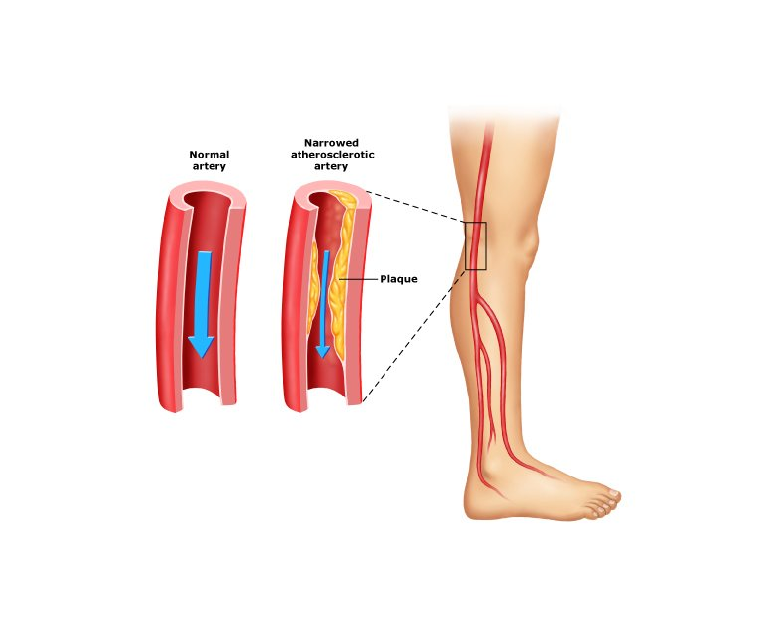
Coronary Artery Disease (CAD)

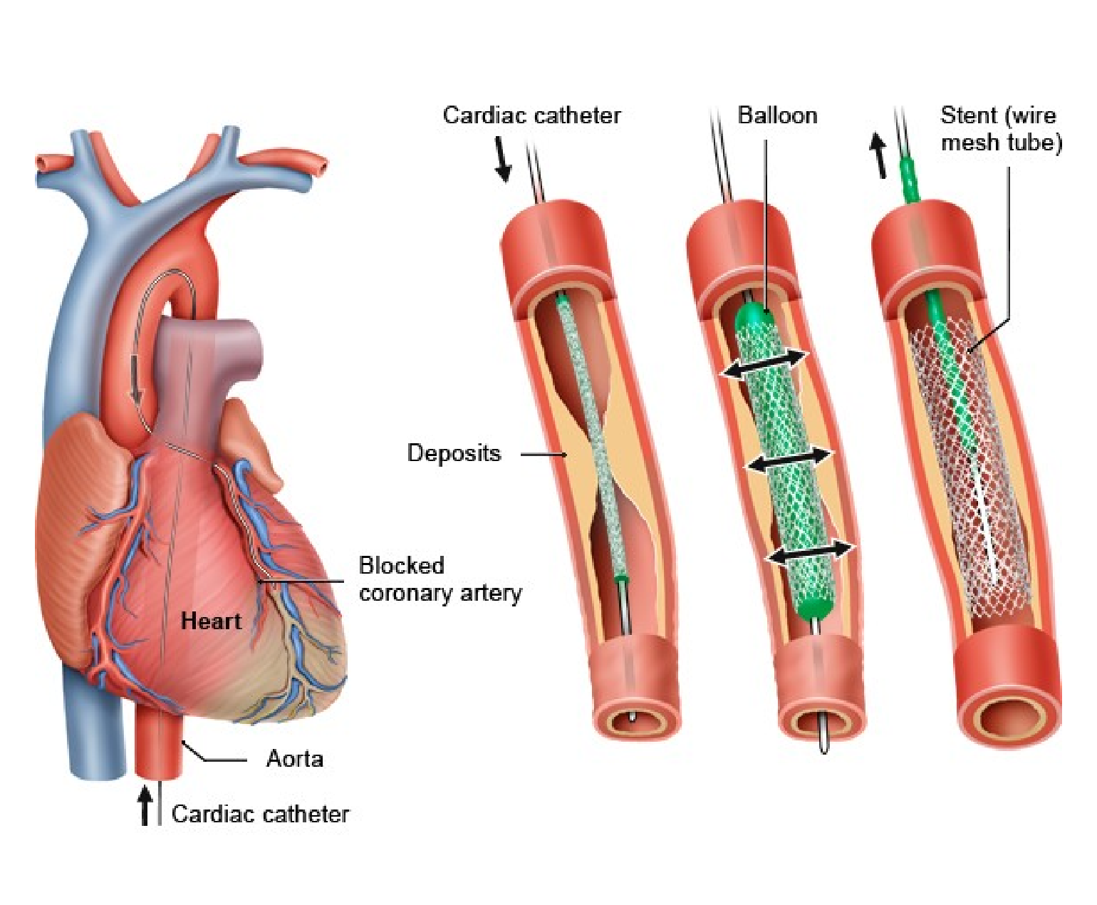
One of the most prevalent cardiovascular conditions in older adults is coronary artery disease (CAD). CAD occurs when the arteries that supply blood to the heart become narrowed or blocked due to atherosclerosis, the build-up of plaque. Older adults are more at risk to CAD due to factors such as prolonged exposure to risk factors like high blood pressure, high cholesterol, and smoking.

Diagnosis of CAD in older patients may be challenging because symptoms can be masked by other age-related conditions. Doctors employ diagnostic tools such as stress tests and cardiac imaging to confirm the diagnosis. Treatment typically involves a combination of lifestyle modifications, medication management, and, in severe cases, invasive interventions like angioplasty or bypass surgery.
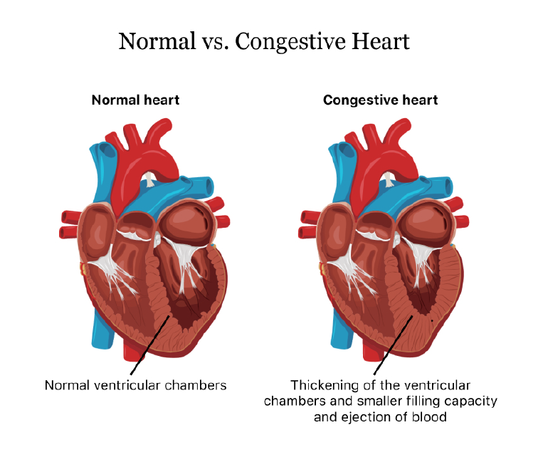
Heart failure

Heart failure is another common cardiovascular condition in older patients. It occurs when the heart is unable to pump blood efficiently, leading to symptoms such as shortness of breath, fatigue, and fluid retention. Age-related changes in the heart, such as reduced elasticity and stiffening of the cardiac muscles, increase the risk of heart failure.

Accurate diagnosis of heart failure requires a comprehensive assessment of symptoms, physical examination, and diagnostic tests like echocardiography. Treatment involves a combination of medications to manage symptoms and improve heart function, lifestyle modifications like fluid and salt restriction, and, in some cases, devices like pacemakers or implantable defibrillators.

Arrhythmias

Arrhythmias, including atrial fibrillation (AF), are commonly encountered in older adults. AF, characterized by irregular and rapid heartbeat, is associated with an increased risk of stroke, heart failure, and other cardiovascular complications.

Advanced age is a significant risk factor for developing AF due to changes in the electrical conduction system of the heart. Diagnosis involves an electrocardiogram (ECG) and may require additional monitoring methods like Holter monitoring or event recorders. Management of AF in older patients aims to control heart rate, prevent blood clots with anticoagulant therapy, and restore normal heart rhythm when possible.

Hypertension

Hypertension, or high blood pressure, is highly prevalent in older adults and is a major risk factor for cardiovascular disease. Age-related changes in blood vessels, hormonal factors, and lifestyle habits contribute to the development of hypertension.

Effective management of hypertension in older patients involves lifestyle modifications, such as a heart-healthy diet and regular exercise, along with medications to achieve blood pressure targets. Close monitoring and regular follow-up are essential to ensure blood pressure control and prevent complications.

Conclusion

Cardiovascular conditions in older patients require a comprehensive and individualized approach to diagnosis and treatment. Age-related changes in the cardiovascular system increase the vulnerability of older adults to conditions like coronary artery disease, heart failure, arrhythmias, and hypertension. Addressing modifiable risk factors and promoting healthy lifestyle habits are essential for preventing and managing cardiovascular conditions in older patients.

Congestive Heart Failure

Congestive heart failure (CHF) is a condition in which the heart is unable to pump blood effectively to meet the body’s needs. This results in a build-up of fluid in the lungs, liver, and other tissues, leading to symptoms such as shortness of breath, fatigue, and swelling in the legs and ankles.

CHF can be caused by a variety of factors, including coronary artery disease, high blood pressure, and valve disorders. It can also occur as a result of certain medical conditions, such as diabetes, obesity, and thyroid disorders. Lifestyle factors such as smoking, excessive alcohol consumption, and lack of exercise can also increase the risk of CHF.

Symptoms

Symptoms of CHF can vary depending on the severity of the condition and the underlying cause. Some of the most common symptoms include:

  1. Shortness of breath: This is often the first symptom of CHF and can occur even during normal activities such as walking or climbing stairs. The shortness of breath can be accompanied by coughing or wheezing.
  2. Fatigue: People with CHF often experience extreme tiredness, even after minimal physical activity.
  3. Swelling: Fluid build-up in the legs, ankles, and feet can cause swelling, also known as edema. This can also occur in the abdomen and other areas of the body.
  4. Rapid heartbeat: CHF can cause the heart to beat faster than normal in an attempt to compensate for the decreased pumping ability.
  5. Decreased appetite: CHF can cause a loss of appetite and nausea, which can lead to weight loss.

If left untreated, CHF can lead to serious complications such as kidney failure, liver damage, and even death. However, with proper treatment and management, many people with CHF can live healthy and active lives.

Diagnosis & Treatment

Diagnosis of CHF typically involves a physical exam, medical history, and diagnostic tests such as chest x-rays, electrocardiograms (ECGs), and echocardiograms. These tests can help identify the underlying cause of the condition and determine the extent of the damage to the heart.

Treatment for CHF typically involves a combination of lifestyle changes, medications, and in some cases, surgical procedures. Some of the most common treatments for CHF include:

  1. Medications: Medications such as diuretics, ACE inhibitors, and beta-blockers can help reduce fluid build-up, lower blood pressure, and improve heart function.
  2. Lifestyle changes: Lifestyle changes such as quitting smoking, reducing alcohol consumption, and maintaining a healthy weight can help improve heart function and reduce the risk of complications.
  3. Cardiac rehabilitation: This is a supervised exercise program designed to help people with CHF improve their fitness levels and overall health.
  4. Implantable devices: Devices such as pacemakers and implantable cardioverter-defibrillators (ICDs) can help regulate the heartbeat and prevent dangerous heart rhythms.
  5. Surgery: In some cases, surgery may be necessary to repair or replace damaged heart valves or to bypass blocked arteries.

Conclusion

While CHF is a serious condition, there are many things that people with the condition can do to improve their overall health and quality of life. This includes consuming a healthy diet, regular exercise, avoiding smoking and excessive alcohol consumption. It is also important for people with CHF to consult their doctor regularly for monitoring of symptoms and medication adjustments as needed. By adopting these measures, people with CHF can manage their symptoms and reduce their risk of complications.

Ross Procedure

The Ross procedure is a surgical procedure used to treat aortic valve disease, which is a condition in which the valve that controls blood flow from the heart to the aorta becomes narrowed or leaks. This procedure involves replacing the damaged aortic valve with the patient’s own pulmonary valve, and then replacing the pulmonary valve with a donated or artificial valve.

The Ross procedure is named after its creator, Donald Ross, a Scottish cardiac surgeon who developed the technique in the 1960s. Since then, the procedure has become a popular option for treating aortic valve disease in certain patients.

Advantages of Ross Procedure

The Ross procedure is typically recommended for younger patients who have aortic valve disease, as it has several advantages over other types of valve replacement surgery. One of the main advantages is that it avoids the need for lifelong anticoagulation therapy, which is required for patients who receive a mechanical valve replacement. Anticoagulation therapy is a type of medication that helps prevent blood clots, but it can also increase the risk of bleeding and other complications.

Another advantage of the Ross procedure is that it provides a better quality of life for patients. The pulmonary valve is better suited to the low-pressure environment of the aortic position than artificial valves, which can often lead to complications such as valve dysfunction or blood clots.

Procedure

The Ross procedure is performed under general anaesthesia and typically takes many hours to complete. During the procedure, the surgeon will make an incision in the chest and access the heart. The patient’s pulmonary valve will then be removed and used to replace the damaged aortic valve. A new pulmonary valve, either from a donated cadaver or an artificial valve, will then be used to replace the removed pulmonary valve.

After the procedure, patients may need to spend several days in the hospital recovering. They may experience some discomfort or pain, and will need to take medication to manage pain and prevent infection. Patients will also need to gradually increase their physical activity and follow a prescribed rehabilitation program to help them recover.

Suitability

While the Ross procedure has many benefits, it is not suitable for all patients with aortic valve disease. Patients who have other heart conditions, such as coronary artery disease or an enlarged heart, may not be good candidates for the procedure. Additionally, patients who have already had a previous valve replacement or who have other medical conditions that could complicate surgery may not be good candidates for the procedure.

Conclusion

Ross procedure is a surgical procedure used to treat aortic valve disease in certain patients. It involves replacing the damaged aortic valve with the patient’s own pulmonary valve, and then replacing the pulmonary valve with a donated or artificial valve. If you are considering the Ross procedure, it is important to discuss the risks and benefits with your doctor to determine if it is the right option for you.

What is Peripheral Artery Disease?

Peripheral artery disease (PAD) is a circulatory condition that affects the peripheral arteries, which are the blood vessels that carry blood from the heart to the arms, legs, and other parts of the body. PAD occurs when these arteries become narrow or blocked, reducing blood flow to the affected body parts. The most common symptoms of PAD include pain, numbness, and cramping in the legs or feet, especially during physical activity.

How is PAD caused?

PAD is caused by atherosclerosis, a condition in which fatty deposits, or plaques, build up inside the arteries, narrowing the blood vessels and restricting blood flow. Atherosclerosis can affect any artery in the body, but it is most commonly found in the peripheral arteries of the legs and feet. Other risk factors for PAD include smoking, high blood pressure, high cholesterol, diabetes, obesity, and a family history of the disease.

PAD can be a serious condition, and if left untreated, it can lead to complications such as foot ulcers, gangrene, and amputation. However, there are a number of treatments available for PAD, and early diagnosis and treatment can help prevent these complications and improve quality of life.

Treatment

The first step in treating PAD is to make lifestyle changes that can help improve blood flow and reduce the risk of further narrowing of the arteries. These may include quitting smoking, losing weight, exercising regularly, and following a healthy diet.

Medications may also be prescribed to help manage symptoms and reduce the risk of complications. Some of the most commonly used medications for PAD include antiplatelet agents, such as aspirin or clopidogrel, which can help prevent blood clots from forming in the narrowed arteries, and statins, which can help lower cholesterol levels and reduce the risk of further plaque build-up.

In more severe cases of PAD, surgical or minimally invasive procedures may be necessary to restore blood flow to the affected areas. These procedures may include angioplasty, in which a small balloon is inserted into the narrowed artery and inflated to widen the artery and improve blood flow, or stenting, in which a small metal mesh tube is placed in the artery to hold it open.

In some cases, bypass surgery may be necessary, in which a healthy blood vessel is taken from another part of the body and used to bypass the blocked artery, allowing blood to flow freely to the affected area.

If left untreated, PAD can lead to serious complications, including foot ulcers, gangrene, and amputation. However, with proper diagnosis and treatment, the prognosis for PAD is generally good. By making lifestyle changes, taking medication as prescribed, and undergoing any necessary procedures, most people with PAD can improve their symptoms, reduce their risk of complications, and maintain their mobility and quality of life.

Conclusion

PAD is a common circulatory condition that can cause pain, numbness, and cramping in the legs and feet, especially during physical activity. It is caused by atherosclerosis, and risk factors include smoking, high blood pressure, high cholesterol, diabetes, obesity, and a family history of the disease. Treatment for PAD may include lifestyle changes, medications, or surgical procedures, depending on the severity of the condition. Early diagnosis and treatment can help prevent complications and improve quality of life for people with PAD.

Truncus Arteriosus

Truncus arteriosus is a rare congenital heart defect that occurs in approximately 1 in 10,000 live births. It is a serious condition that requires prompt medical attention and intervention to prevent potentially life-threatening complications.

What is Truncus arteriosus?

In a normal heart, the blood is pumped from the right side of the heart to the lungs, where it is oxygenated, and then returned to the left side of the heart, where it is pumped out to the rest of the body. This process is facilitated by two separate arteries, the pulmonary artery, which carries blood to the lungs, and the aorta, which carries blood to the rest of the body.

However, in the case of truncus arteriosus, the pulmonary artery and the aorta fail to separate during foetal development, resulting in a single large vessel that arises from both the right and left ventricles. This vessel is known as the truncus arteriosus. Additionally, there is usually a hole in the wall that separates the ventricles, called a ventricular septal defect (VSD).

As a result of this defect, oxygen-rich and oxygen-poor blood mix together in the truncus arteriosus before being pumped out to the body. This results in a lack of oxygen to the body’s tissues, leading to symptoms such as fatigue, shortness of breath, and poor growth.

Truncus arteriosus can also lead to more serious complications such as heart failure, pulmonary hypertension, and developmental delays. The severity of the condition depends on the size of the VSD and the degree of mixing of blood within the truncus arteriosus.

Diagnosis & Treatment

Truncus arteriosus is typically diagnosed soon after birth or during infancy, with symptoms such as a bluish tint to the skin (cyanosis), rapid breathing, and poor feeding. Diagnostic tests such as echocardiography, cardiac catheterization, and magnetic resonance imaging (MRI) may be performed to confirm the diagnosis and evaluate the severity of the defect.

Treatment for truncus arteriosus usually involves surgery to repair the defect. This surgery typically involves dividing the truncus arteriosus into two separate vessels, the pulmonary artery and the aorta, and patching the VSD. The timing of the surgery depends on the severity of the defect, but it is typically performed within the first few months of life.

Following surgery, regular follow-up appointments with a pediatric cardiologist are necessary to monitor the child’s progress and ensure that the repair is successful. Some children may require additional procedures or treatments, such as medication for pulmonary hypertension or developmental therapy for delays.

The prognosis for children with truncus arteriosus depends on the severity of the defect and the presence of any associated conditions. However, with prompt diagnosis, early intervention, and appropriate medical care, many children with truncus arteriosus go on to lead healthy, active lives.

Conclusion

Truncus arteriosus is a rare but serious congenital heart defect that requires prompt medical attention and intervention. Parents and caregivers should be aware of the symptoms and seek medical attention if they suspect their child may have this condition. With appropriate treatment and follow-up care, many children with truncus arteriosus can lead healthy, fulfilling lives.

What is Cardiac Catheterization?

Cardiac catheterization, also known as coronary angiography, is a medical procedure that allows doctors to examine the heart and blood vessels to identify any problems. It is a minimally invasive procedure that involves inserting a thin, flexible tube called a catheter into a blood vessel, typically in the groin or arm, and guiding it through the blood vessels to the heart.

The catheterization procedure is performed in a cardiac catheterization laboratory (also known as a cath lab) by a cardiologist. The procedure is typically done under local anaesthesia, which numbs the area where the catheter will be inserted.

Procedure

During the procedure, the cardiologist will make a small incision in the skin and insert a hollow sheath into the blood vessel. The catheter is then inserted through the sheath and guided to the heart using x-ray guidance. Once the catheter is in place, the cardiologist can inject a contrast dye into the bloodstream, which makes the heart and blood vessels visible on x-ray.

The contrast dye allows the cardiologist to examine the heart and blood vessels for any abnormalities or blockages that may be present. If a blockage is found, the cardiologist can perform a procedure called angioplasty to open the blocked artery and restore blood flow to the heart.

Diagnostic Tool

Cardiac catheterization is a valuable diagnostic tool that can provide important information about the heart and blood vessels. It is typically used to diagnose and evaluate a range of heart conditions, including coronary artery disease, heart valve problems, congenital heart defects, and heart failure.

In addition to providing diagnostic information, cardiac catheterization can also be used to treat certain heart conditions. For example, angioplasty can be used to open blocked arteries and restore blood flow to the heart. Other procedures, such as stent placement and balloon valvuloplasty, can also be performed during cardiac catheterization.

Risks

While cardiac catheterization is generally considered a safe procedure, there are some risks associated with it. These include bleeding, infection, damage to the blood vessels, and allergic reactions to the contrast dye. However, these risks are generally low and can be minimized with proper preparation and monitoring.

Tests before the procedure

Before undergoing cardiac catheterization, patients will typically undergo a series of tests to evaluate their heart health and determine if they are a good candidate for the procedure. These tests may include an electrocardiogram (ECG), echocardiogram, stress test, and blood tests.

Patients should also inform their doctor of any medications they are taking, as some medications may need to be temporarily stopped prior to the procedure. Patients should also avoid eating or drinking anything for several hours before the procedure.

Conclusion

Overall, cardiac catheterization is a valuable tool for diagnosing and treating a range of heart conditions. While there are some risks associated with the procedure, these risks are generally low and can be minimized with proper preparation and monitoring. Patients should talk to their doctor about whether cardiac catheterization is a good option for them and what they can expect during and after the procedure.

Electrical Cardioversion

Electrical cardioversion is a medical procedure that is used to treat abnormal heart rhythms, also known as arrhythmias. This procedure involves the delivery of a controlled electric shock to the heart through two large pads or paddles placed on the chest. The shock interrupts the abnormal electrical activity of the heart and allows it to reset to its normal rhythm.

Arrhythmias

Arrhythmias can occur in the upper chambers of the heart (atria) or the lower chambers (ventricles). The most common arrhythmias are atrial fibrillation, atrial flutter, and supraventricular tachycardia. These arrhythmias can cause symptoms such as palpitations, dizziness, shortness of breath, chest pain, and fatigue. If left untreated, they can also increase the risk of stroke, heart failure, and other serious complications.

Electrical cardioversion procedure

Electrical cardioversion is performed by a trained healthcare provider such as a cardiologist. Before the procedure, the patient is given medications to reduce the risk of blood clots and to ensure that the heart is in the correct rhythm. The cardiologist then places the pads or paddles on the chest of the patient, and a brief electric shock is delivered to the heart.

The amount of electricity delivered is carefully calibrated to ensure that it is enough to reset the heart’s rhythm without causing any damage to the heart or surrounding tissues. The shock can cause the patient’s muscles to contract, which can sometimes be painful. However, patients are typically given sedation or anaesthesia to minimize discomfort during the procedure.

Once the heart has been reset to its normal rhythm, the patient is monitored closely for a period of time to ensure that the heart remains stable. In some cases, additional electrical shocks may be needed to maintain the normal rhythm.

Electrical cardioversion is a highly effective procedure for treating certain types of arrhythmias. In many cases, it can restore normal heart function almost immediately. However, the success of the procedure depends on several factors, including the type and severity of the arrhythmia, the duration of the arrhythmia, and the overall health of the patient.

Medical evaluation before electrical cardioversion

Before undergoing electrical cardioversion, patients will usually undergo a thorough medical evaluation to ensure that they are good candidates for the procedure. This evaluation may include blood tests, an electrocardiogram (ECG), a chest X-ray, and other tests as needed. The healthcare provider will also review the patient’s medical history, including any medications or supplements that the patient is taking.

Patients with certain conditions may not be suited for electrical cardioversion. For example, patients with certain types of heart disease, severe lung disease, or a history of blood clots may be at increased risk of complications from the procedure. The doctor will carefully evaluate the patient’s medical history and perform any necessary tests to determine whether electrical cardioversion is safe and appropriate.

Electrical cardioversion is generally a safe procedure, but like any medical procedure, it can have some risks. The most common side effect is mild skin irritation or burns where the pads or paddles are placed on the chest. These typically heal within a few days.

Patients may also be advised to make certain lifestyle changes, such as quitting smoking, reducing alcohol intake, and losing weight, to reduce the risk of future arrhythmias.

Conclusion

Electrical cardioversion is an effective treatment for certain types of arrhythmias. With proper care and follow-up, many patients can expect to have improved heart function and reduced risk of future arrhythmias.

Complete Atrioventricular Canal Defect (CAVC)

Complete atrioventricular canal defect (CAVC) is a rare congenital heart condition that occurs when there is a hole in the centre of the heart that affects the atria and ventricles. This condition is also known as an atrioventricular septal defect (AVSD), and it can cause a range of serious health problems if left untreated.

In a normal heart, there is a wall (septum) between the two atria and another wall between the two ventricles. These walls prevent the mixing of oxygen-rich and oxygen-poor blood. In CAVC, these walls are missing, and a large hole exists between the atria and ventricles. As a result, oxygen-rich blood mixes with oxygen-poor blood, leading to reduced oxygen levels in the body and heart failure over time.

Diagnosis of CAVC

CAVC is typically diagnosed in infancy or early childhood. Infants with CAVC may experience symptoms such as difficulty breathing, rapid breathing, and feeding difficulties. In some cases, babies with CAVC may also have low oxygen levels, which can cause a bluish tint to the skin (cyanosis).

The diagnosis of CAVC is usually confirmed through diagnostic tests such as an echocardiogram, which uses ultrasound waves to create images of the heart. Other tests that may be performed include an electrocardiogram (ECG) to evaluate the heart’s electrical activity and a chest X-ray to check for signs of heart enlargement.

Treatment

Treatment for CAVC usually involves surgery to close the hole in the heart and reconstruct the affected valves. This type of surgery is typically done in early infancy or early childhood, depending on the severity of the condition. In some cases, medication may be prescribed to manage symptoms such as heart failure or to prevent complications such as infection.

Long-term outlook

The long-term outlook for children with CAVC depends on the severity of the defect and the timing of treatment. In general, children who receive timely and appropriate treatment have a good prognosis and can live normal, healthy lives. However, in severe cases, CAVC can lead to heart failure, pulmonary hypertension, and other serious complications.

In addition to medical treatment, children with CAVC may require ongoing monitoring and care throughout their lives. This may include regular check-ups with a pediatric cardiologist, medication management, and follow-up diagnostic tests such as echocardiograms.

Conclusion

Complete atrioventricular canal defect is a rare but serious congenital heart condition that affects the atria and ventricles of the heart. Early diagnosis and appropriate treatment are crucial for managing the condition and preventing complications. If you suspect your child may have CAVC, it is important to seek medical attention promptly to ensure the best possible outcome.

How Positron Emission Tomography (PET) Scan Works?

Positron emission tomography (PET) is a medical imaging technique that produces detailed images of the inside of the body. It is used to diagnose and monitor various medical conditions, including cancer, heart disease, and neurological disorders.

What is a PET scan?

A PET scan is a type of medical imaging that uses a small amount of a radioactive substance, called a tracer, to produce images of the inside of the body. The tracer is injected into the body, swallowed, or inhaled, and then detected by a PET scanner. The PET scanner is a large machine that contains a ring of detectors that can detect the radiation emitted by the tracer.

How does a PET scan work?

The tracer used in a PET scan is typically a compound that contains a small amount of a radioactive substance, such as fluorine-18 or carbon-11. These radioactive substances emit positrons, which are positively charged particles that are the opposite of electrons. When a positron encounters an electron in the body, they annihilate each other and emit two gamma rays in opposite directions.

The PET scanner detects the gamma rays and uses them to create a three-dimensional image of the area being scanned. The scanner can detect the gamma rays from multiple angles, which allows it to create a detailed image of the area.

What happens during a PET scan?

Before the PET scan, the patient is injected with the tracer. The tracer is typically injected into a vein in the arm, but it can also be swallowed or inhaled, depending on the type of scan being done.

After the tracer is injected, the patient is asked to rest for a period of time to allow the tracer to circulate through the body. The amount of time the patient needs to rest depends on the type of scan being done and the tracer being used.

Once the tracer has circulated through the body, the patient is taken to the PET scanner. The scanner is a large machine with a narrow table that slides in and out of the machine.

The patient lies on the table, and the scanner is positioned over the area being scanned. The patient needs to remain still during the scan to ensure the images are clear.

The scanner emits a small amount of radiation to detect the tracer in the body. The patient will not feel anything during the scan, but they may hear a clicking or buzzing sound coming from the machine.

After the scan is complete, the patient can go home. The amount of radiation in the tracer is small, and it typically does not pose a health risk.

What are the uses of PET scans?

PET scans are used to diagnose and monitor a variety of medical conditions, including:

  1. Cancer: PET scans can help to diagnose cancer and determine the extent of the disease. They can also be used to monitor the effectiveness of cancer treatments, such as chemotherapy.
  2. Heart disease: PET scans can be used to diagnose heart disease and determine the extent of damage to the heart muscle. They can also be used to assess blood flow to the heart and to determine the effectiveness of treatments, such as angioplasty.
  3. Neurological disorders: PET scans can be used to diagnose and monitor neurological disorders, such as Alzheimer’s disease, Parkinson’s disease, and epilepsy.
  4. Mental health disorders: PET scans can be used to study the brain and to diagnose and monitor mental health disorders, such as depression and schizophrenia.

What is Vasculitis?

Vasculitis is a medical condition that affects the blood vessels in the body, leading to inflammation and damage to these vital structures. The blood vessels affected by vasculitis can range from small capillaries to larger arteries, depending on the type of vasculitis. This damage to the blood vessels can cause a range of symptoms and health problems, including impaired blood flow, organ damage, and increased risk of infections and bleeding.

Types of Vasculitis

There are several different types of vasculitis, each with its own unique causes, symptoms, and treatment options. Some of the most common types of vasculitis include giant cell arteritis, Takayasu’s arteritis, and Wegener’s granulomatosis. These diseases can affect people of all ages, and can be caused by a variety of factors, including infections, autoimmune disorders, and exposure to certain medications or chemicals.

Symptoms

The symptoms of vasculitis can vary widely, depending on the type and severity of the disease, as well as the location of the affected blood vessels. Some common symptoms of vasculitis include fever, fatigue, weight loss, joint pain, skin rashes, and sores that do not heal. In severe cases, vasculitis can also cause organ damage, such as kidney failure, heart attack, or stroke.

Diagnosis and Treatment

Diagnosing vasculitis can be challenging, as the symptoms can be similar to other diseases and conditions. The process usually involves a thorough medical history, physical examination, blood tests, and imaging studies, such as MRI or CT scans. In some cases, a biopsy may be necessary to confirm the diagnosis.

Treatment for vasculitis depends on the type and severity of the disease, as well as the age and overall health of the patient. In many cases, treatment involves a combination of medications, such as corticosteroids and immunosuppressants, to reduce inflammation and prevent further damage to the blood vessels. These medications can be taken orally or intravenously, and may need to be taken for an extended period of time.

In severe cases of vasculitis, treatment may also involve other interventions, such as stents, surgery or radiation therapy. For example, if the vasculitis is causing a blockage in a major blood vessel, such as the aorta, surgery may be necessary to remove the blockage and restore normal blood flow.

Living with vasculitis

Living with vasculitis can be challenging, but with proper treatment and care, many people are able to manage their symptoms and lead healthy lives. It is important to consult a specialist and monitor your symptoms and medical condition regularly. This may include regular blood tests, imaging studies, and physical exams, as well as lifestyle changes, such as maintaining a healthy diet and getting regular exercise.

Conclusion

Vasculitis is a group of rare diseases characterized by inflammation and damage to the blood vessels in the body. The symptoms and treatment options can vary widely depending on the type and severity of the disease, but with proper care and management, many people are able to live active, healthy lives.

Posts navigation