Preoperative and Postoperative Care

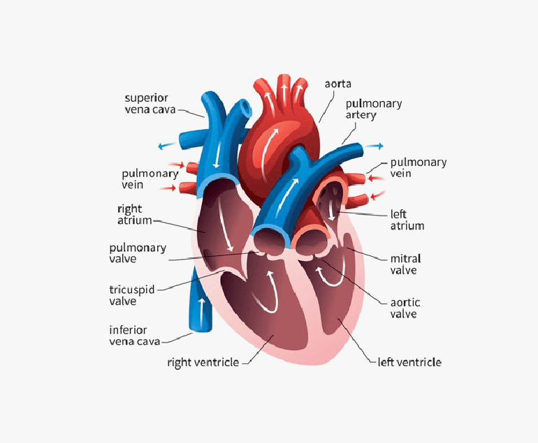
The preoperative and postoperative care for Coronary Artery Bypass Grafting (CABG) is crucial for the overall success of the procedure and the patient’s recovery. Here’s an overview of the care provided before and after CABG:

Preoperative Care:

  1. Medical Evaluation:
    1. A thorough medical history and physical examination are conducted to assess the patient’s overall health and identify potential risks.
    1. Diagnostic tests, such as electrocardiogram (ECG), echocardiogram, and blood tests, help evaluate the extent and severity of coronary artery disease.
  2. Cardiac Catheterization and Angiography:
    1. Coronary angiography is often performed to visualize the coronary arteries and determine the location and severity of blockages.
  3. Medication Management:
    1. Medications, such as antiplatelets and anticoagulants, may be prescribed to manage blood clotting and reduce the risk of complications.
  4. Smoking Cessation:
    1. Patients are strongly encouraged to quit smoking before surgery to improve their overall cardiovascular health.
  5. Dietary Guidelines:
    1. Dietary modifications may be recommended to manage cholesterol levels and optimize the patient’s nutritional status.
  6. Preoperative Education:
    1. Patients receive information about the surgery, potential risks, and postoperative care.
    1. Rehabilitation plans and lifestyle changes are discussed.

Postoperative Care:

  1. Immediate Postoperative Period (ICU):
    1. Patients are closely monitored in the intensive care unit (ICU) initially.
    1. Vital signs, fluid balance, and cardiac function are closely observed.
  2. Pain Management:
    1. Adequate pain control is essential for patient comfort and to facilitate early mobility.
    1. Pain medications are administered as needed.
  3. Monitoring and Assessment:
    1. The continuous monitoring of heart rate, blood pressure, oxygen saturation, and other vital signs is necessary.
    1. Regular assessments of wound sites, neurological status, and respiratory function.
  4. Respiratory Care:
    1. Breathing exercises and incentive spirometry are encouraged to prevent complications like atelectasis and pneumonia.
    1. Early ambulation helps improve lung function.
  5. Medication Management:
    1. Medications, including antiplatelets, anticoagulants, and medications to control blood pressure and cholesterol, are prescribed as needed.
  6. Nutritional Support:
    1. Nutritional needs are assessed, and a plan is developed to ensure the patient receives adequate nourishment for recovery.
  7. Rehabilitation and Physical Therapy:
    1. Gradual mobilization and physical therapy are initiated to improve strength and endurance.
    1. Cardiac rehabilitation programs may be recommended for long-term cardiovascular health.
  8. Follow-up and Lifestyle Changes:
    1. To monitor progress, it is necessary to have regular follow-up appointments with healthcare providers.
    1. Implementation of lifestyle changes, including a heart-healthy diet, regular exercise, and smoking cessation.
  9. Emotional Support:
    1. Psychological support is provided to help patients cope with the emotional aspects of surgery and recovery.

Both preoperative and postoperative care are collaborative efforts involving the surgical team, nurses, rehabilitation specialists, and the patient. The goal is to optimize the patient’s overall health, minimize complications, and support a successful recovery. Individual care plans may vary, and patients should follow the specific recommendations provided by their healthcare team.

Both preoperative and postoperative care are collaborative efforts involving the surgical team, nurses, rehabilitation specialists, and the patient. The goal is to optimize the patient’s overall health, minimize complications, and support a successful recovery. Individual care plans may vary, and patients should follow the specific recommendations provided by their healthcare team.

Rheumatic Heart Disease

Rheumatic heart disease (RHD) is a serious condition that affects the heart valves and is a result of complications from rheumatic fever. Although it is preventable and treatable, RHD continues to be a significant health burden in many developing countries, particularly among children and young adults.

Causes

RHD is caused by an abnormal immune response to untreated or inadequately treated streptococcal throat infections, mainly caused by group A streptococcus bacteria. When these infections are not promptly treated with antibiotics, the body’s immune system can mistakenly attack its own tissues, including the heart valves. Over time, this can lead to scarring, thickening, and deformation of the valves, which impairs their ability to function properly.

Symptoms

The symptoms of RHD can vary depending on the severity and location of valve damage. In the early stages, there may be no noticeable symptoms, but as the disease progresses, individuals may experience shortness of breath, chest pain, fatigue, palpitations, and swollen ankles or feet. In severe cases, heart failure and life-threatening complications, such as infective endocarditis or stroke, can occur.

Diagnosis & Treatment

Diagnosing RHD typically involves a combination of medical history assessment, physical examination, echocardiography (ultrasound of the heart), electrocardiogram (ECG), and other imaging tests. These tests help evaluate the structure and function of the heart, identify valve abnormalities, and determine the extent of damage.

Treatment for RHD aims to manage symptoms, prevent disease progression, and reduce the risk of complications. Antibiotic prophylaxis is crucial to prevent recurrent streptococcal infections, which can trigger disease exacerbation. Medications, such as diuretics, beta-blockers, and anticoagulants, may be prescribed to manage symptoms and reduce the risk of blood clots.

Prevention

Preventing RHD begins with prompt and appropriate treatment of streptococcal throat infections. Administering antibiotics, such as penicillin, to individuals with streptococcal infections can prevent the development of rheumatic fever, thereby reducing the risk of RHD. Regular follow-up and secondary prophylaxis, which involves long-term antibiotic treatment, are essential for individuals who had rheumatic fever to prevent disease recurrence and progression.

Conclusion

Rheumatic heart disease is a preventable and treatable condition that continues to be a significant health concern in many parts of the world. Prompt and appropriate treatment of streptococcal throat infections, along with regular follow-up and secondary prophylaxis, are vital for preventing the development and progression of RHD. By prioritizing prevention, early detection, and comprehensive management, the impact of rheumatic heart disease can be minimised.

Electrical Cardioversion

Electrical cardioversion is a medical procedure that is used to treat abnormal heart rhythms, also known as arrhythmias. This procedure involves the delivery of a controlled electric shock to the heart through two large pads or paddles placed on the chest. The shock interrupts the abnormal electrical activity of the heart and allows it to reset to its normal rhythm.

Arrhythmias

Arrhythmias can occur in the upper chambers of the heart (atria) or the lower chambers (ventricles). The most common arrhythmias are atrial fibrillation, atrial flutter, and supraventricular tachycardia. These arrhythmias can cause symptoms such as palpitations, dizziness, shortness of breath, chest pain, and fatigue. If left untreated, they can also increase the risk of stroke, heart failure, and other serious complications.

Electrical cardioversion procedure

Electrical cardioversion is performed by a trained healthcare provider such as a cardiologist. Before the procedure, the patient is given medications to reduce the risk of blood clots and to ensure that the heart is in the correct rhythm. The cardiologist then places the pads or paddles on the chest of the patient, and a brief electric shock is delivered to the heart.

The amount of electricity delivered is carefully calibrated to ensure that it is enough to reset the heart’s rhythm without causing any damage to the heart or surrounding tissues. The shock can cause the patient’s muscles to contract, which can sometimes be painful. However, patients are typically given sedation or anaesthesia to minimize discomfort during the procedure.

Once the heart has been reset to its normal rhythm, the patient is monitored closely for a period of time to ensure that the heart remains stable. In some cases, additional electrical shocks may be needed to maintain the normal rhythm.

Electrical cardioversion is a highly effective procedure for treating certain types of arrhythmias. In many cases, it can restore normal heart function almost immediately. However, the success of the procedure depends on several factors, including the type and severity of the arrhythmia, the duration of the arrhythmia, and the overall health of the patient.

Medical evaluation before electrical cardioversion

Before undergoing electrical cardioversion, patients will usually undergo a thorough medical evaluation to ensure that they are good candidates for the procedure. This evaluation may include blood tests, an electrocardiogram (ECG), a chest X-ray, and other tests as needed. The healthcare provider will also review the patient’s medical history, including any medications or supplements that the patient is taking.

Patients with certain conditions may not be suited for electrical cardioversion. For example, patients with certain types of heart disease, severe lung disease, or a history of blood clots may be at increased risk of complications from the procedure. The doctor will carefully evaluate the patient’s medical history and perform any necessary tests to determine whether electrical cardioversion is safe and appropriate.

Electrical cardioversion is generally a safe procedure, but like any medical procedure, it can have some risks. The most common side effect is mild skin irritation or burns where the pads or paddles are placed on the chest. These typically heal within a few days.

Patients may also be advised to make certain lifestyle changes, such as quitting smoking, reducing alcohol intake, and losing weight, to reduce the risk of future arrhythmias.

Conclusion

Electrical cardioversion is an effective treatment for certain types of arrhythmias. With proper care and follow-up, many patients can expect to have improved heart function and reduced risk of future arrhythmias.

What is Pulmonary Valve Stenosis?

Pulmonary valve stenosis is a medical condition in which the pulmonary valve, which regulates blood flow from the right ventricle of the heart to the lungs, becomes narrowed or obstructed. This can lead to difficulty breathing and reduced oxygen levels in the blood, causing a range of symptoms and potentially serious health complications if left untreated.

Causes of Pulmonary Valve Stenosis

There are several potential causes of pulmonary valve stenosis. One common cause is congenital heart defects, which are present at birth and may be inherited or caused by environmental factors during pregnancy. Rheumatic fever, a condition that can develop as a complication of strep throat, can also lead to pulmonary valve stenosis. In rare cases, the valve may become narrowed or obstructed due to scarring or inflammation. In some cases, the cause of the stenosis is unknown.

Symptoms

Symptoms of pulmonary valve stenosis can vary depending on the severity of the condition. In mild cases, individuals may not experience any symptoms or may only have mild shortness of breath during physical activity. More severe cases may cause significant difficulty breathing, chest pain, fatigue, and fainting. In severe cases, the condition can also cause heart palpitations, swelling in the legs and ankles, and a bluish tint to the skin due to low oxygen levels.

Diagnosis and Treatment

Diagnosis of pulmonary valve stenosis typically begins with a physical exam and a review of the individual’s medical history. The doctor may also order several tests to confirm the diagnosis and assess the severity of the condition. These tests may include an echocardiogram, a chest X-ray, and an electrocardiogram.

There are several treatment options available for pulmonary valve stenosis, depending on the severity of the condition and the individual’s overall health. In mild cases, treatment may include medications to manage symptoms and reduce the risk of complications. These may include diuretics to reduce fluid build-up in the body and beta blockers to reduce the heart’s workload.

For more severe cases, surgery may be necessary to repair or replace the valve. Repair surgery involves making changes to the existing valve to improve its function, while valve replacement surgery involves replacing the faulty valve with a new one. There are several different types of valves that can be used in replacement surgery, including mechanical valves, which are made of man-made materials, and biological valves, which are made from animal tissue.

In some cases, a procedure called balloon valvuloplasty may be used to widen the narrowed valve. This procedure involves inserting a balloon catheter into the narrowed valve and inflating the balloon to widen the opening.

It is important for patients with pulmonary valve stenosis to work closely with their doctors to manage their condition and reduce the risk of complications. This may include regular check-ups and monitoring of symptoms, lifestyle changes such as quitting smoking and eating a healthy diet, and taking prescribed medications as directed.

Conclusion

Pulmonary valve stenosis is a serious condition that can have a significant impact on an individual’s quality of life. With proper treatment and management, however, it is possible to live a full and active life. It is important for individuals with the condition to stay in close communication with their doctors and follow their treatment plan to ensure the best possible outcomes.